Copyright
©The Author(s) 2025.
World J Radiol. Jun 28, 2025; 17(6): 107776
Published online Jun 28, 2025. doi: 10.4329/wjr.v17.i6.107776
Published online Jun 28, 2025. doi: 10.4329/wjr.v17.i6.107776
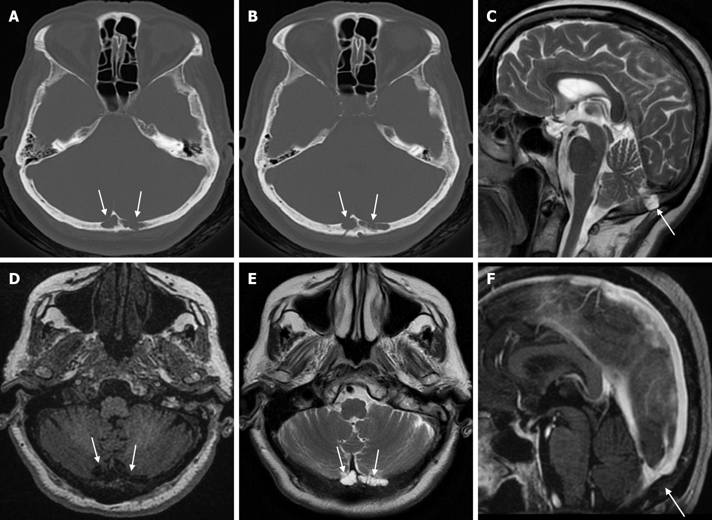
Figure 1 Arachnoid granulations in seventy-eight years old female patient who underwent imaging for headache.
A and B: Axial computed tomography on the bone window show adjacent hypoattenuating structures (arrows) accompanied by cortical destruction in places in the inner table at the level of the internal occipital protuberance of the occipital bone; C: On sagittal T2-weighted image; D: On axial T1-weighted image; E: On axial T2-weighted image; F: On sagittal contrast enhanced T1-weighted image, signal intensities of non-contrast enhanced arachnoid granulation (arrows) are seen, more prominent to the left of the midline in the occipital bone, isointense with cerebrospinal fluid.
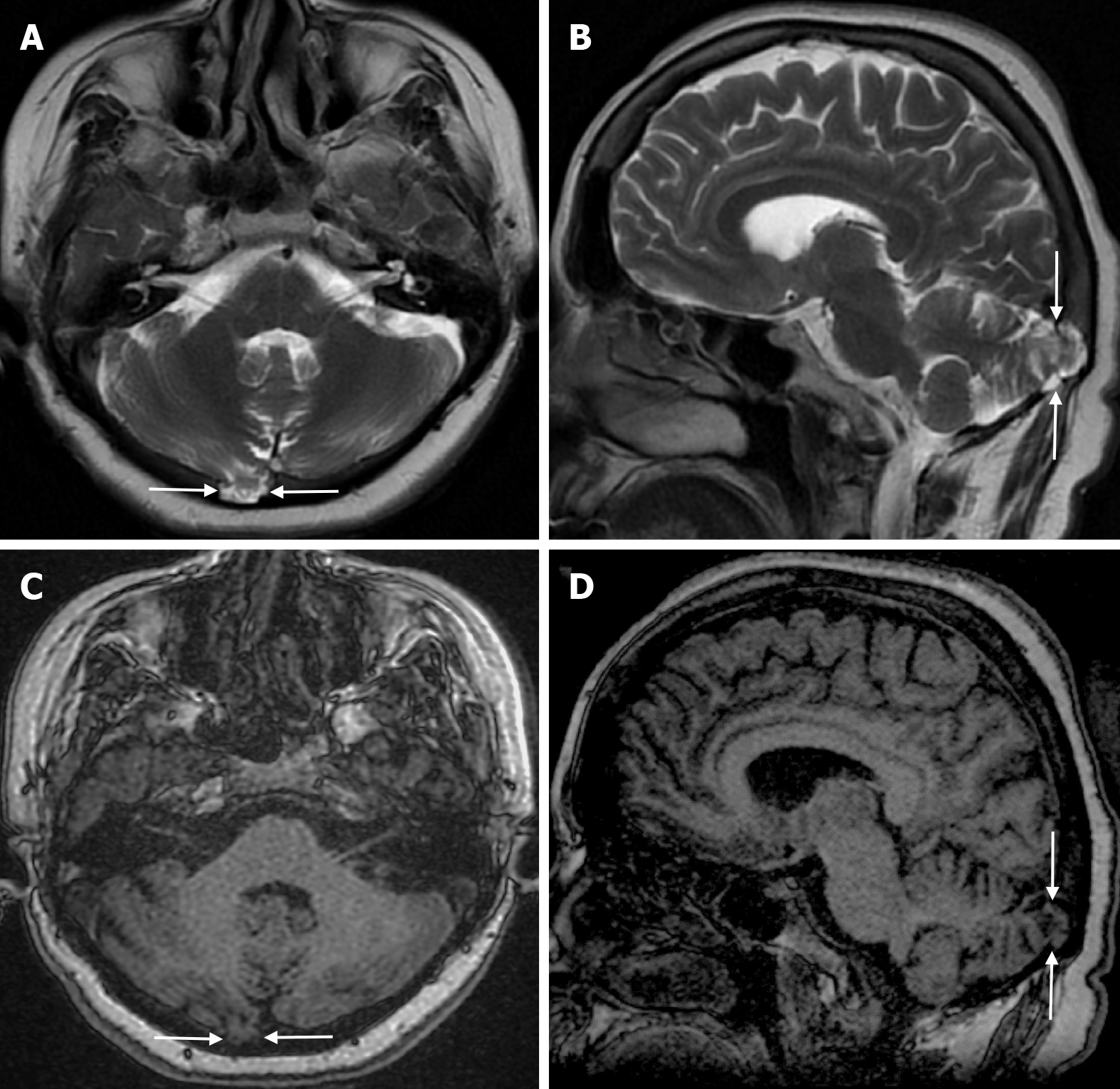
Figure 2 Cerebellar parenchyma herniations into the arachnoid granulations fifty-four-year-old female patient with no history of trauma who underwent imaging for headache.
A: On axial T2-weighted image; B: On sagittal T2-weighted image; C: On axial T1-weighted image; D: On sagittal T1-weighted image, the defect with a maximum diameter of 1.5 centimeter in the inner table to the right of the midline in the right occipital bone and signal intensities of the adjacent cerebellar parenchyma and arachnoid granulation herniating into the diploic space are shown (arrows).
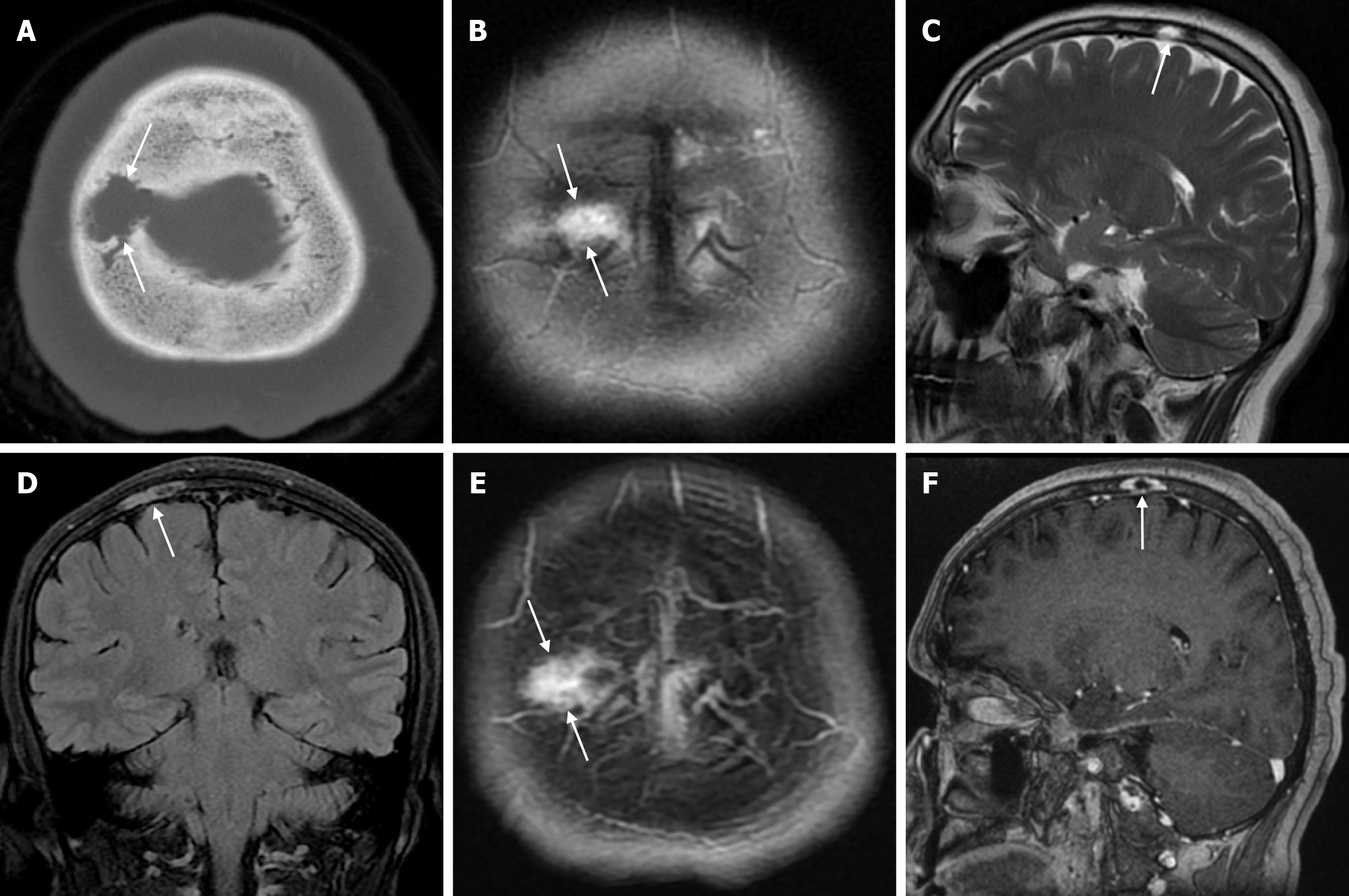
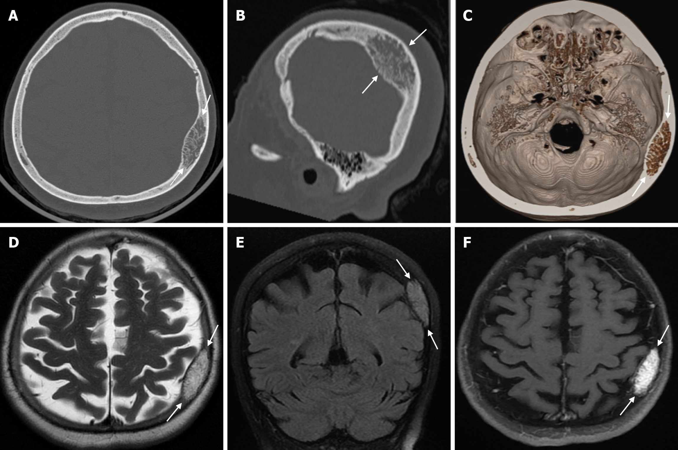
Figure 3 Venous lake in fifty-four years old female patient who underwent imaging for headache.
A; Axial computed tomography on the bone window shows irregular circumscribed structures (arrows) extending to the diploe distance in the right parietal bone in the plane of convexity, without destruction of the outer table; B: On axial T2-weighted image; C: On sagittal T2-weighted image; D: On coronal FLAIR T2-weighted image; E: On axial contrast enhanced T1-weighted image; F: On sagittal contrast enhanced T1-weighted image, in the parasagittal right parietal bone in the plane of convexity, hyperintense on T2-weighted series, hypointense on T1-weighted series, contrast enhanced venous lake (arrows) located in the diploe distance with mild focal irregularity in the inner table. FLAIR: Fluid-attenuated inversion recovery.
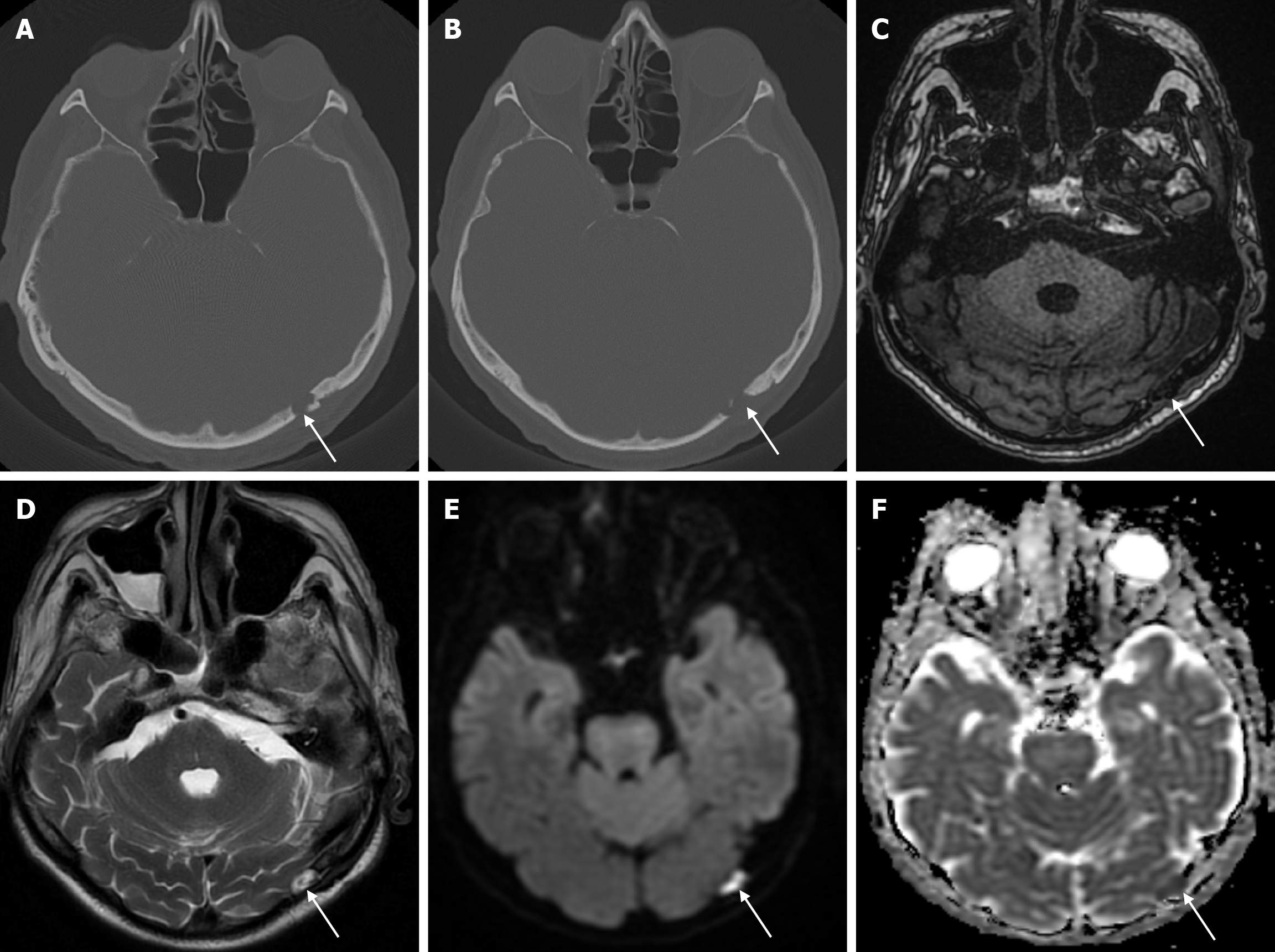
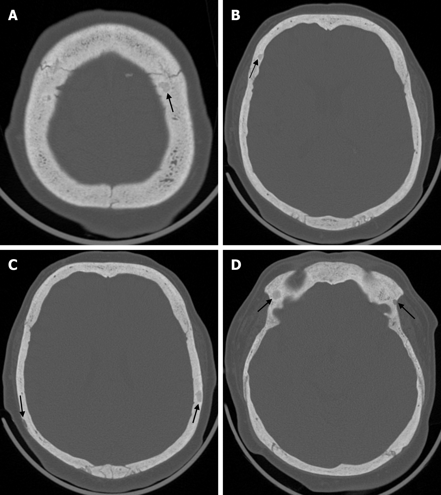
Figure 4 Epidermoid cyst in sixty-five years old male patient who underwent imaging for syncope.
A and B: Axial computed tomography on the bone window show a lesion (arrows) in the left occipital bone adjacent to the lamboid suture with an irregularity in the outer table; C: On axial T1-weighted image, the well-circumscribed lesion in the left occipital bone is hypointense; D: On axial T2-weighted image, the lesion is hyperintense; E: On axial diffusion-weighted image, the lesion is hyperintense; F: On axial apparent diffusion coefficient image, the lesion is hypointense and shows diffusion restriction (arrows).
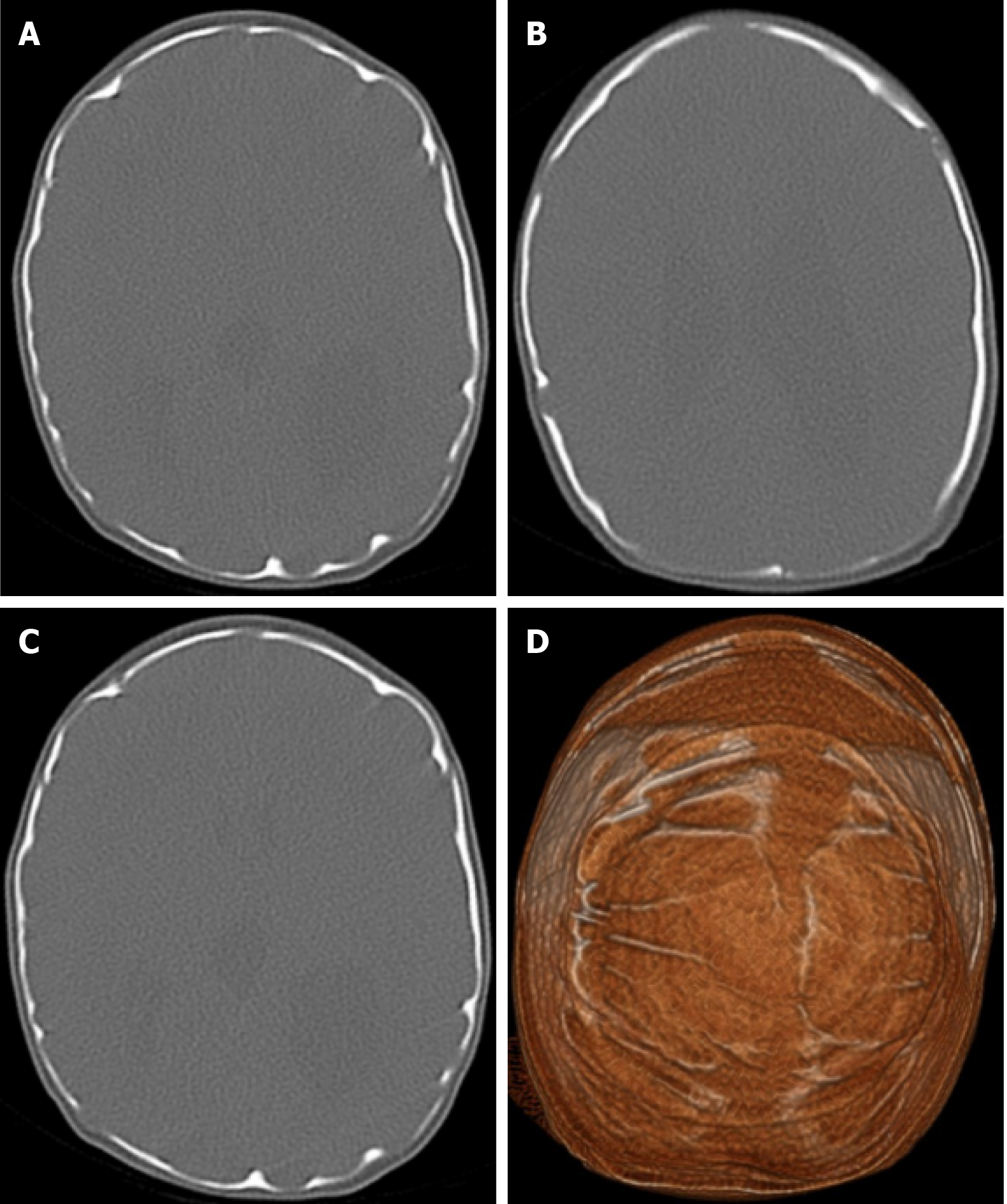
Figure 5 Beaten copper skull of ten days old baby girl undergoing imaging for hydrocephalus.
A, B and C; Axial computed tomography on the bone window; D: On three-dimensional computed tomography scan shows a beaten copper skull with marked curvature of the calvarial bones.
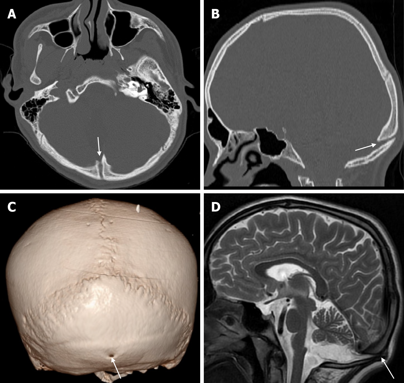
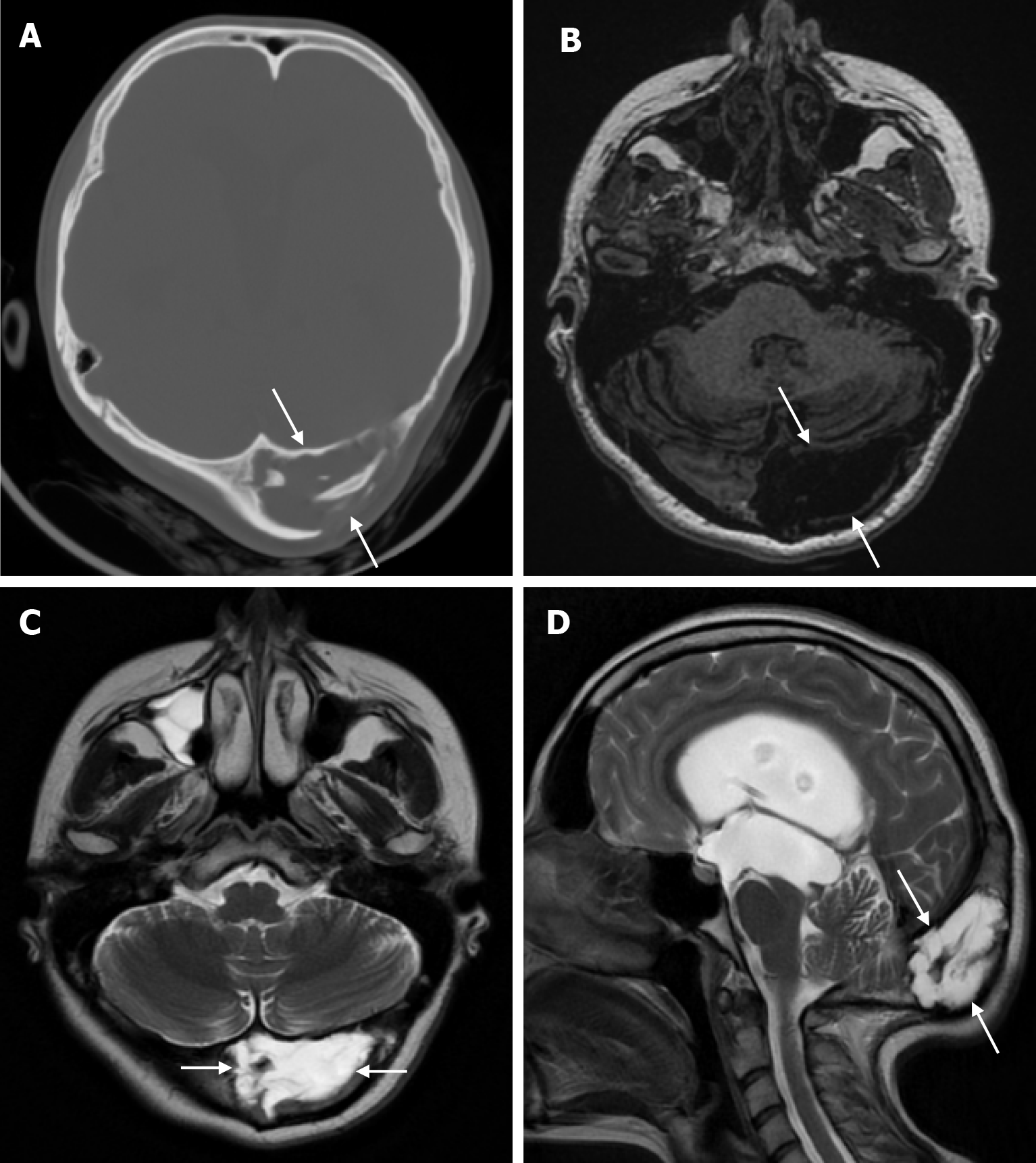
Figure 6 Atretic encephalocele in sixteen years old male patient who underwent imaging for epilepsy.
A and B: Computed tomography on the bone window; C: On three-dimensional computed tomography; D: On sagittal T2-weighted image shows the defective appearance of atretic encephalocele sequelae in the occipital bone in the midline (arrows).
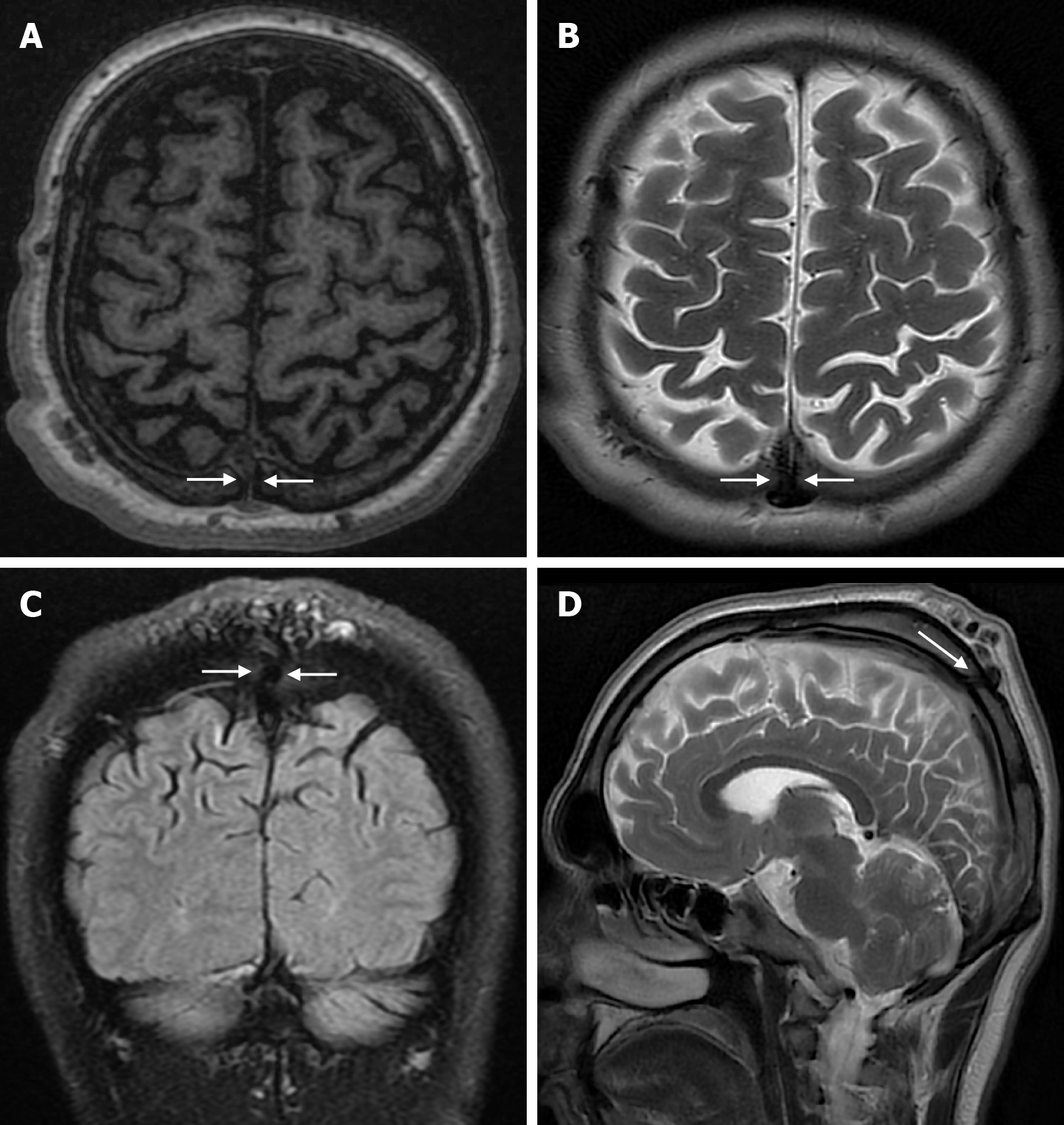
Figure 7 Sinus pericranii in twenty years old male patient who underwent imaging for headache.
A: On axial T1-weighted image; B: On axial T2-weighted image; C: On coronal FLAIR T2-weighted image; D: On sagittal T2-weighted image shows a sinus pericranii associated with the superior sagittal sinus leading to enlarged tortuous venous structures extending to the scalp with a transcarvalial venous duct (arrows) in the midline parietal bone.
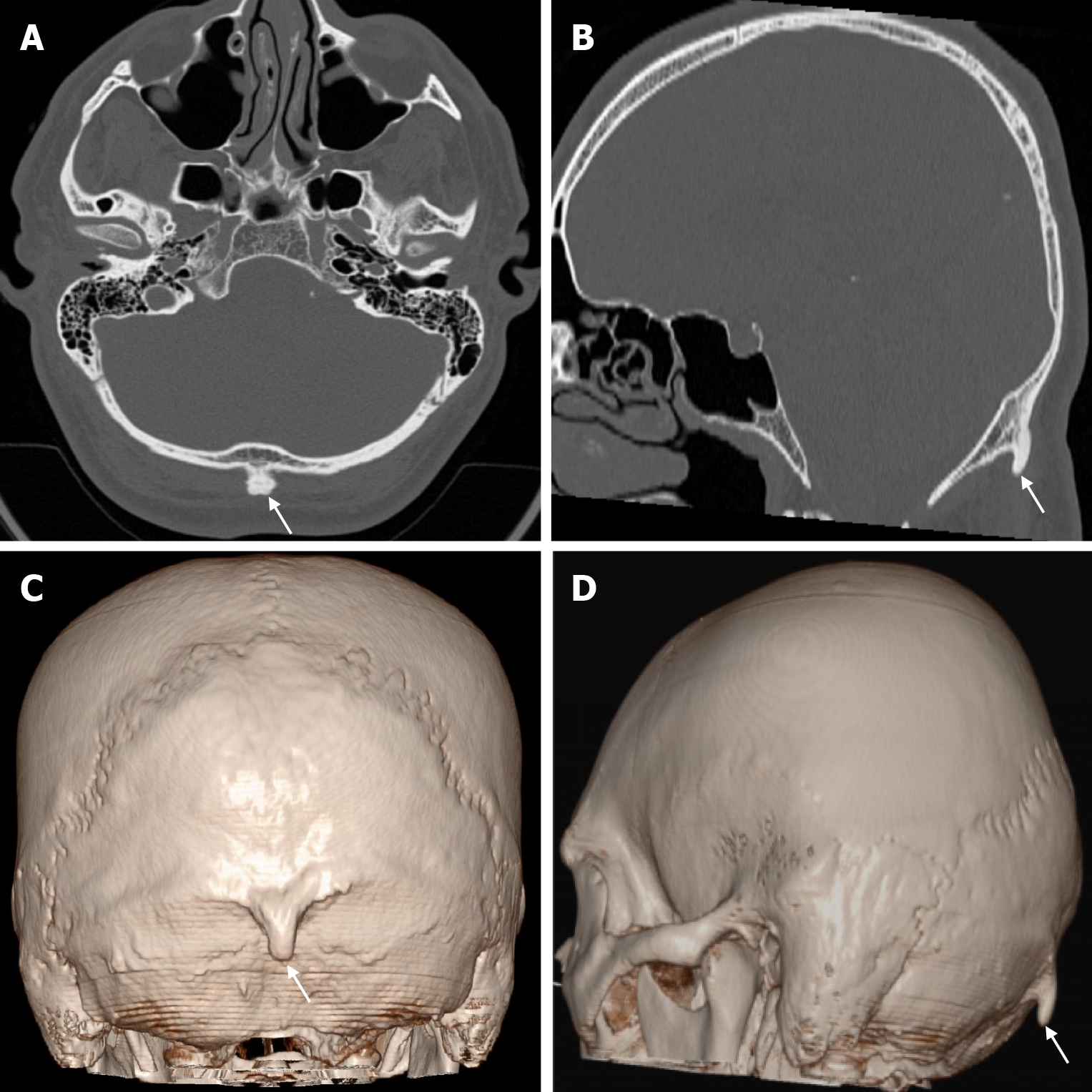
Figure 8 External occipital protuberance (type III) in twenty-nine years old male patient who underwent imaging for headache.
A and B: Computed tomography on the bone window show a bony protuberance (arrows) posterior to the occipital bone; C and D: Three-dimensional computed tomography show an occipital spur (arrows) posterior to the occipital bone.
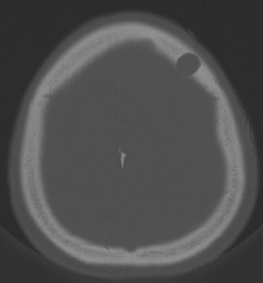
Figure 9 Burr hole in seventy-two years old male patient who underwent surgery for intracranial hemorrhage 1 month ago.
Axial computed tomography on the bone window shows a round calvarial defect in the left frontal bone.
Figure 10 Leptomeningeal cyst in twenty years old female patient who underwent imaging due to a history of trauma at the age of one year.
A: Axial computed tomography on the bone window shows expansion and fluid values (arrows) in the left occipital bone; B: On axial T1-weighted image; C: On axial T2-weighted image; D: Sagittal T2-weighted image shows an isointense leptomeningeal cyst (arrows) with cerebrospinal fluid extending into the left occipital bone and expanding the bone.
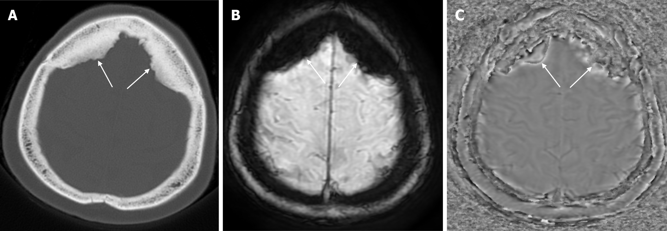
Figure 11 Hyperostosis frontalis interna in sixty-seven years old female patient who underwent imaging for headache.
A: Axial computed tomography on the bone window shows symmetrical calvarial thickening (arrows) of similar density to the bone extending from the inner table to the extra-axial space in the bilateral frontal bones; B: Hyperostosis frontalis interna is hypointense on axial enhanced susceptibility weighted angiography (eSWAN) images, C: Hyperintense on axial filtered phase eSWAN images, signal intensities compatible with hyperosteosis frontalis interna (arrows) at the level of the inner table of the frontal bone.
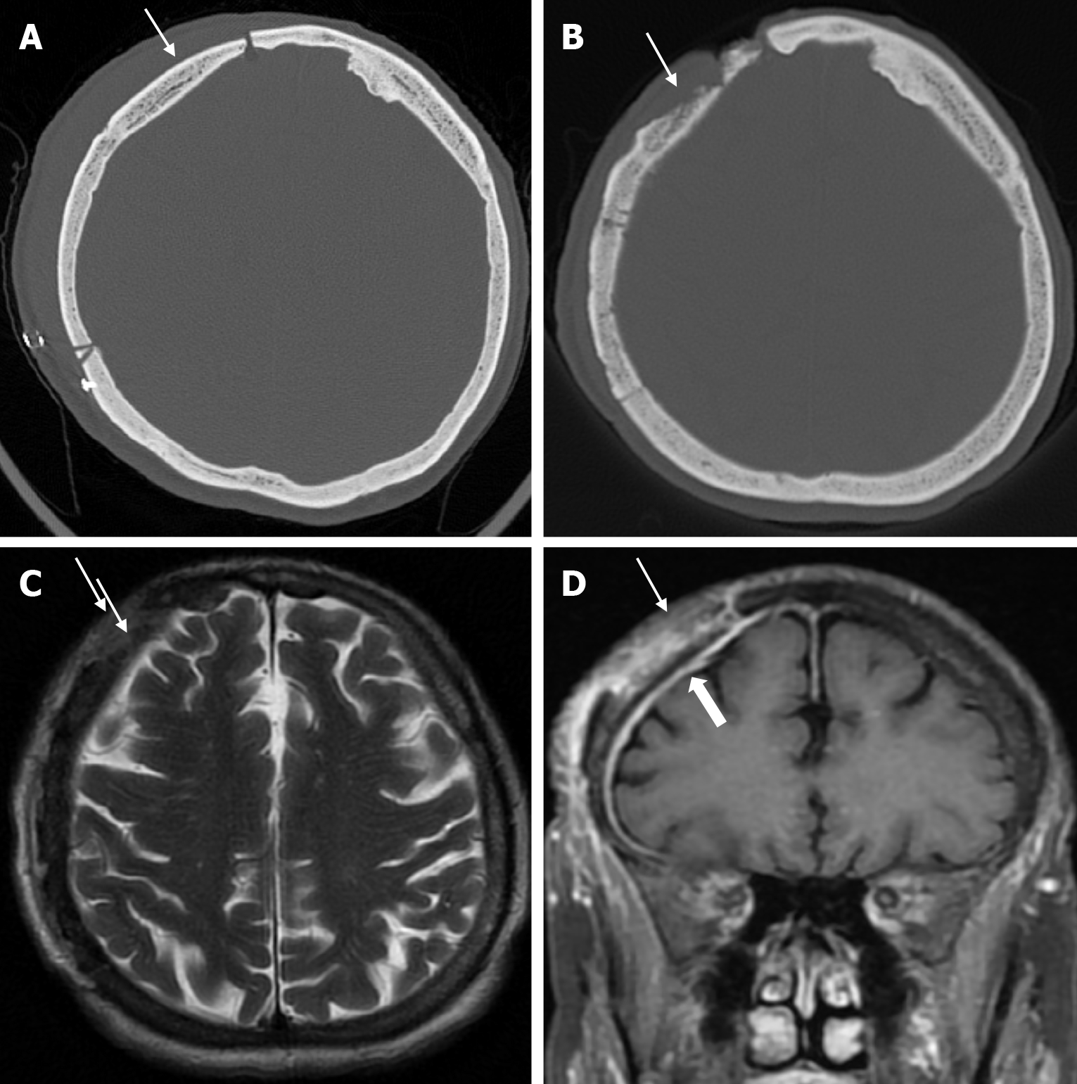
Figure 12 Calvarial osteomyelitis in sixty-nine years old female who underwent surgery for a subdural hematoma.
A: Axial computed tomography of the bone window shows a defect in the right frontoparietal bone secondary to the previous surgery and the outer calvarial table (thin arrow) is intact; B: Axial computed tomography of the bone window six months later shows irregularity and thinning of the outer table (thin arrow) at the surgical site; C: Axial T2-weighted image shows hyperintensity (thin arrow) in the frontoparietal bone; D: On coronal contrast enhanced T1-weighted image, irregularities in the frontoparietal cortical surfaces and heterogeneous contrast enhancement in the bone (thin arrow) and contrast enhancement in the adjacent dura (thick arrow).
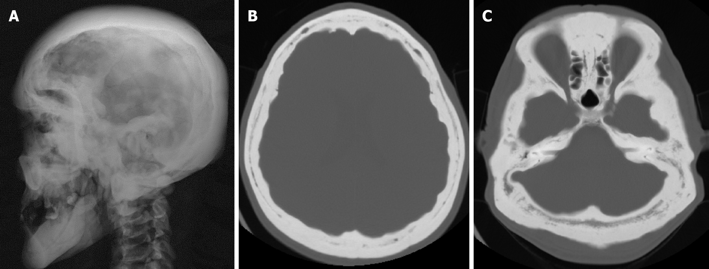
Figure 13 Paget’s disease in thirty-five years old male patient who underwent imaging because of headaches.
A: On lateral plain radiography; A and B: Axial computed tomography of the bone window shows thickening and diffuse sclerosis in all calvarial bones.
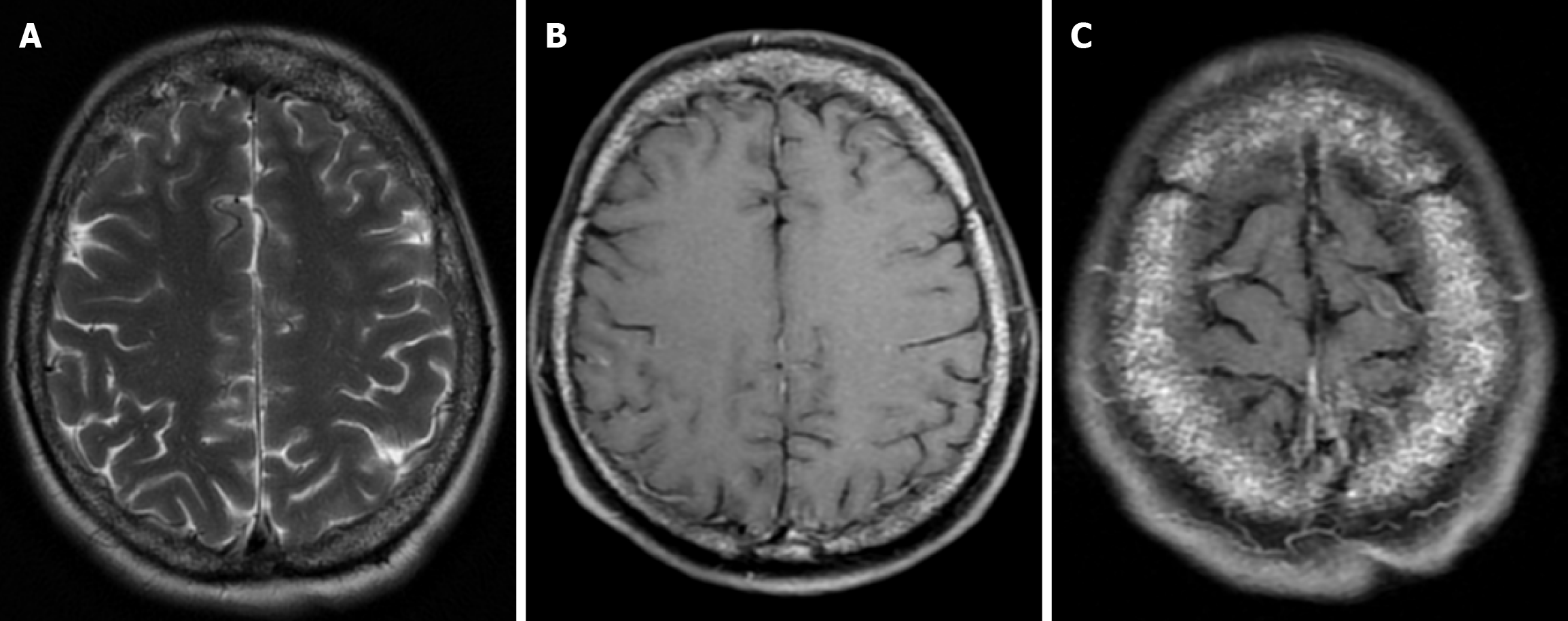
Figure 14 Thalassemia major with calvarial involvement in forty-three years old female patient.
A: On axial T2-weighted image, the skull bones show heterogeneous signal intensity at diploe distance; B and C: On axial fat-saturated contrast enhanced T1-weighted image, the skull bones show bone marrow lesions secondary to thalassaemia major with diffuse heterogeneous contrast enhancement.
Figure 15 Renal osteodystrophy in thirty years old male patient with chronic renal insufficiency who underwent imaging due to confu
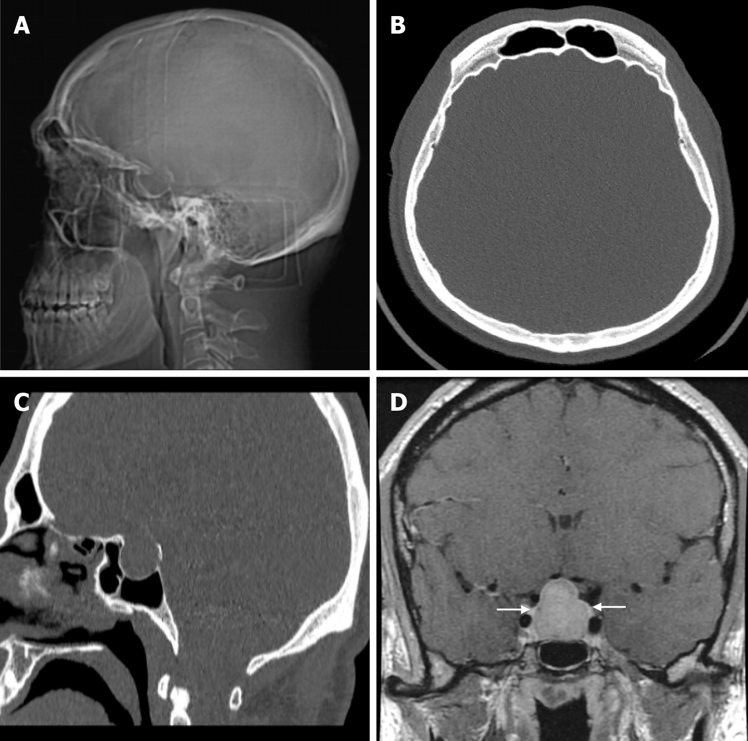
Figure 16 Acromegaly in thirty-two years old male patient who underwent imaging for gigantism.
A: Plain radiography shows thickening of all calvarial bones and expansion of the sella cavity; B: Axial computed tomography on the bone window shows thickening of all calvarial bones; C: Sagittal computed tomography on the bone window shows expansion of the sella cavity; D: Coronal contrast enhanced T1-weighted image shows contrast enhancement macroadenoma (arrows) with smooth lobulated contour completely filling and enlarging the sella cavity, obliterating the suprasellar cistern and compressing the optic chiasm.
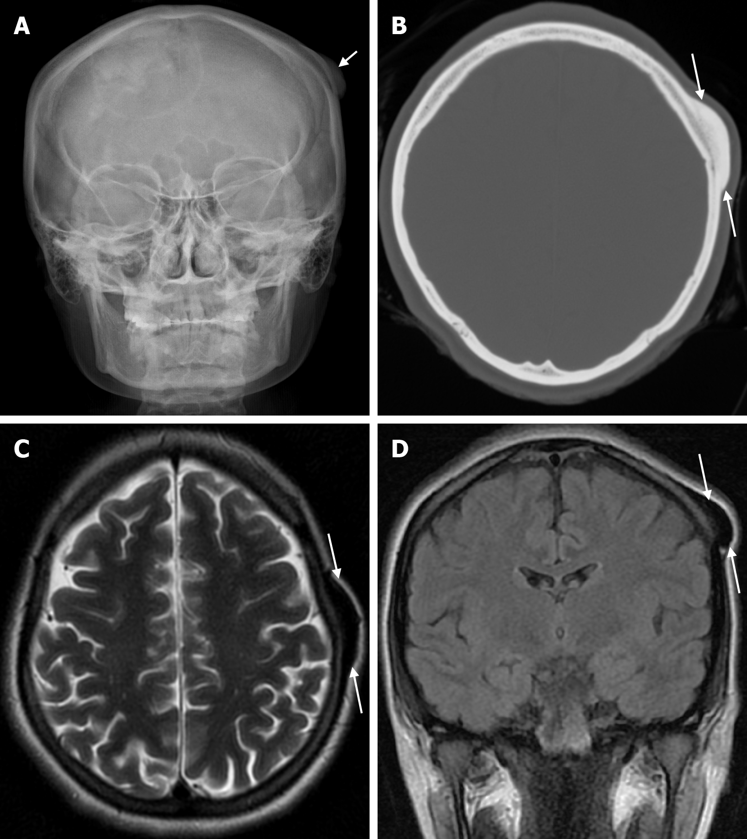
Figure 17 Osteoma in twenty-five years old female patient who underwent imaging for swelling of the scalp.
A: Plain anteroposterior head radiography shows a smooth contoured lesion (arrow) in a compact bony structure extending from the calvarial outer table; B: Axial computed tomography on the bone window shows an isodense smooth contoured lesion (arrows) with broad-based bony structures extending from the outer table to the scalp in the parietal bone; C and D: The left parietal bone shows a smooth contoured sclerotic lesion (arrows) with protrusion from the outer table to the scalp, with a prominent hypointense signal on T2-weighted images.
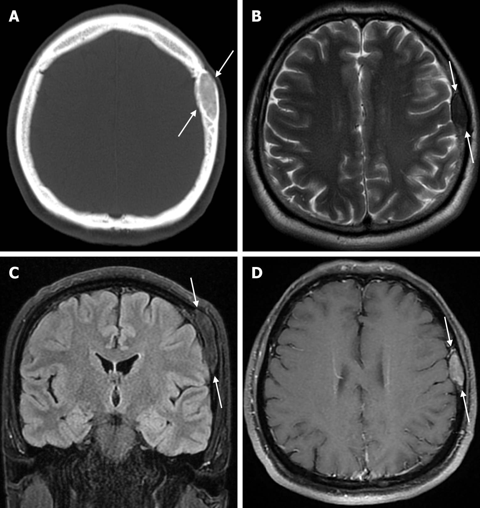
Figure 18 Fibrous dysplasia in thirty-five years old male patient who underwent imaging for headache.
A: Axial computed tomography on the bone window shows a smooth contoured lesion with ground glass density (arrows) in the left parietal bone; B: On axial T2-weighted image, hypointense lesion in the parietal bone (arrows); C: On coronal FLAIR T2-weighted image, mild hyperintense lesion in the parietal bone (arrows); D: Axial contrast enhanced T1-weighted image shows a well-circumscribed lesion (arrows) with contrast enhancement in the parietal bone. FLAIR: Fluid-attenuated inversion recovery.
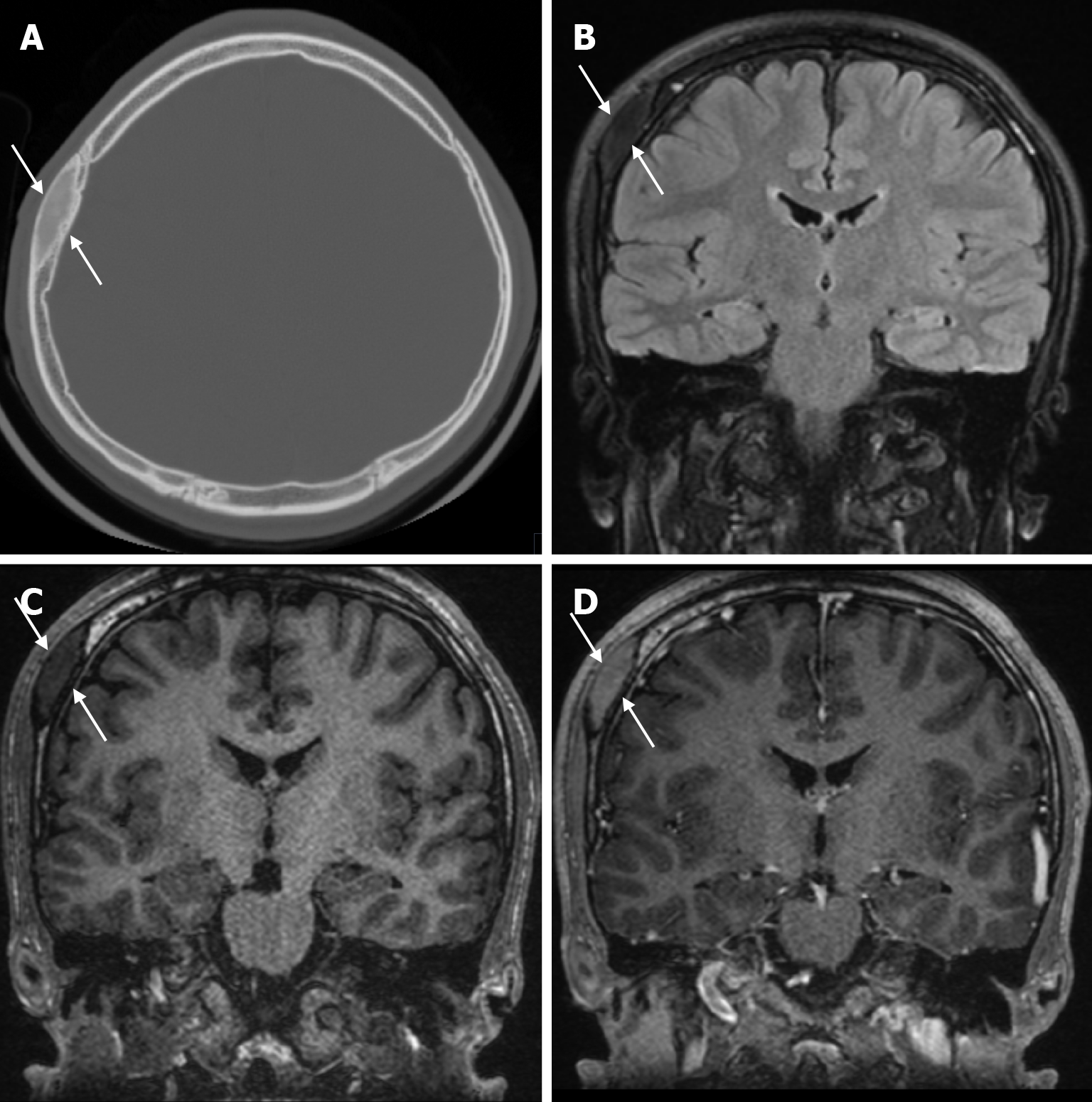
Figure 19 Fibrous dysplasia in twenty years old male patient who underwent imaging for headache.
A: Axial computed tomography on the bone window, the right parietal bone shows a smooth contoured fibrous dysplasia (arrows) close to the bone density, indented to the outer table, causing a slight extension of the diploe, mimicking an osteoma; B: On coronal FLAIR T2-weighted image, mild heterogeneous hypointense lesion in the parietal bone (arrows); C: On coronal T1-weighted image, mild heterogeneous hypointense lesion in the parietal bone (arrows); D: On coronal contrast enhanced T1-weighted image shows contrast enhancement well-circumscribed lesion (arrows) in the parietal bone.
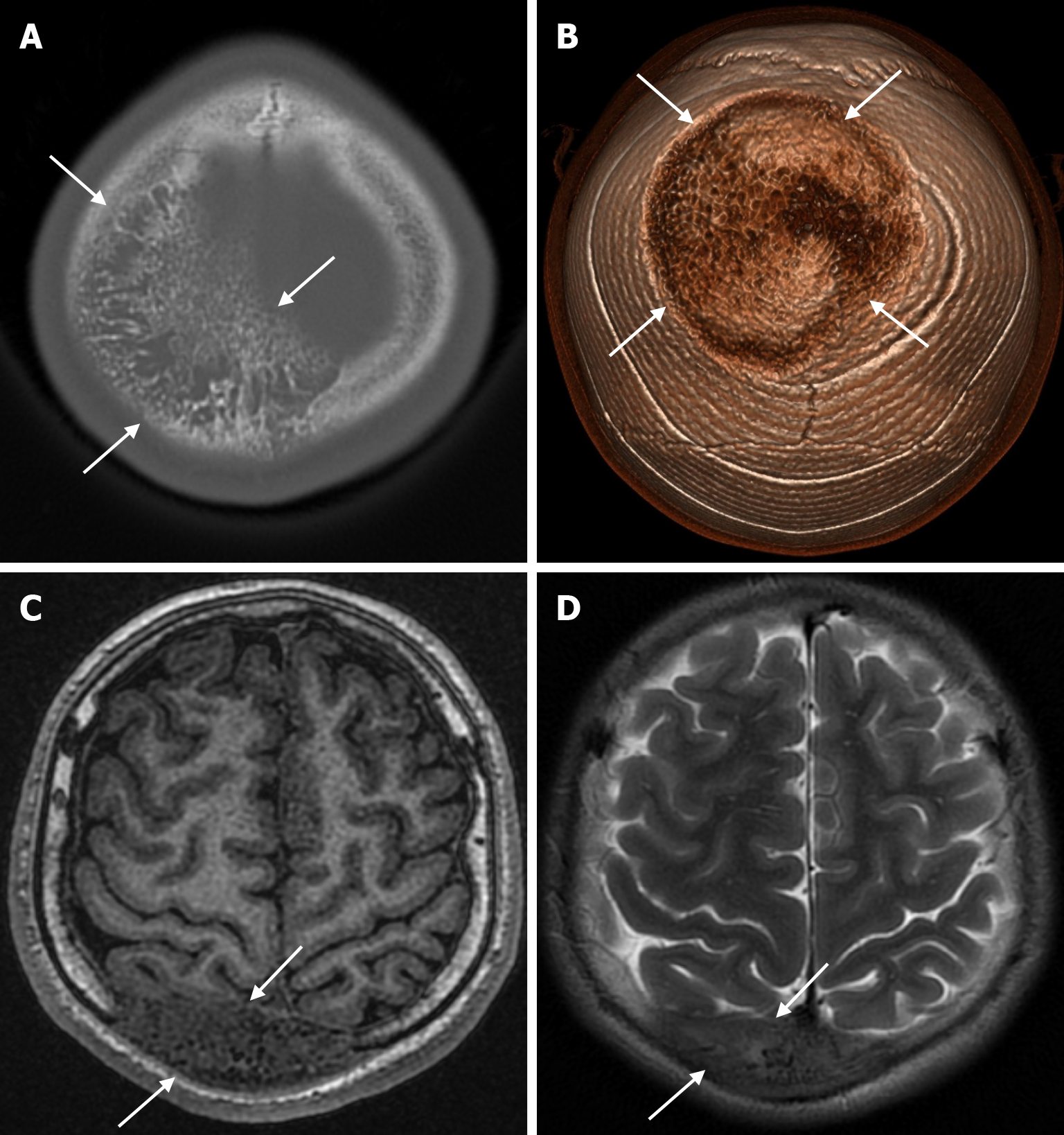
Figure 20 Fibrous dysplasia in seventeen years old male patient who underwent imaging for a calvarial mass.
A: Axial computed tomography on the bone window shows fibrous dysplasia (arrows) mimicking intraosseous venous malformation in both parietal bones, predominantly involving the right parietal bone in the plane of convexity, with coarse trabecular bone structures and lytic areas in a radial pattern involving the inner and outer table and protruding beyond the bone; B: Three-dimensional computed tomography shows fibrous dysplasia protruding from both parietal bones (arrows); C: On axial T1-weighted image, the lesion is heterogeneously hypointense in both parietal bones (arrows); D: Axial T2-weighted image shows heterogeneous mild hyperintensity (arrows) in both parietal bones.
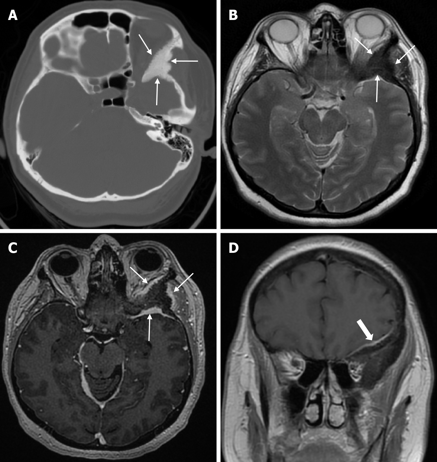
Figure 21 Primary intraosseous meningioma in forty-nine years old female patient who underwent imaging for headache.
A: Axial computed tomography on the bone window shows a smooth-contoured lesion (thin arrows) involving the posterosuperolateral wall of the left orbit, extending to the sphenoid and temporal bones, causing bone expansion and sclerosis, and extending beyond the bone; B: On axial T2-weighted image, the lesion is heterogeneously hypointense (thin arrows); C: On axial contrast enhanced T1-weighted image, the lesion shows cortical-dominated radial irregularities and mild contrast enhancement of bone with a soft tissue component in the vicinity (thin arrows); D: On coronal contrast enhanced T1-weighted image shows contrast enhancement of the dural surface (thick arrow) adjacent to the lesion.
Figure 22 Intraosseous venous malformation in seventy-one years old female patient who underwent imaging for headache.
A and B: Sagittal computed tomography on the bone window show a lytic lesion (arrows) in the left parietal bone, localized in the diploe region, causing widening of the inner table, in which bone trabeculae can be intensely selected; C: Three-dimensional computed tomography shows an intraosseous venous malformation (arrows) in the left parietal bone; D: The lesion is hyperintense (arrows) on axial T2-weighted image; E: On coronal fluid-attenuated inversion recovery T2-weighted image the lesion is hyperintense (arrows); F: On axial contrast enhanced T1-weighted image the lesion shows intense contrast enhancement (arrows).
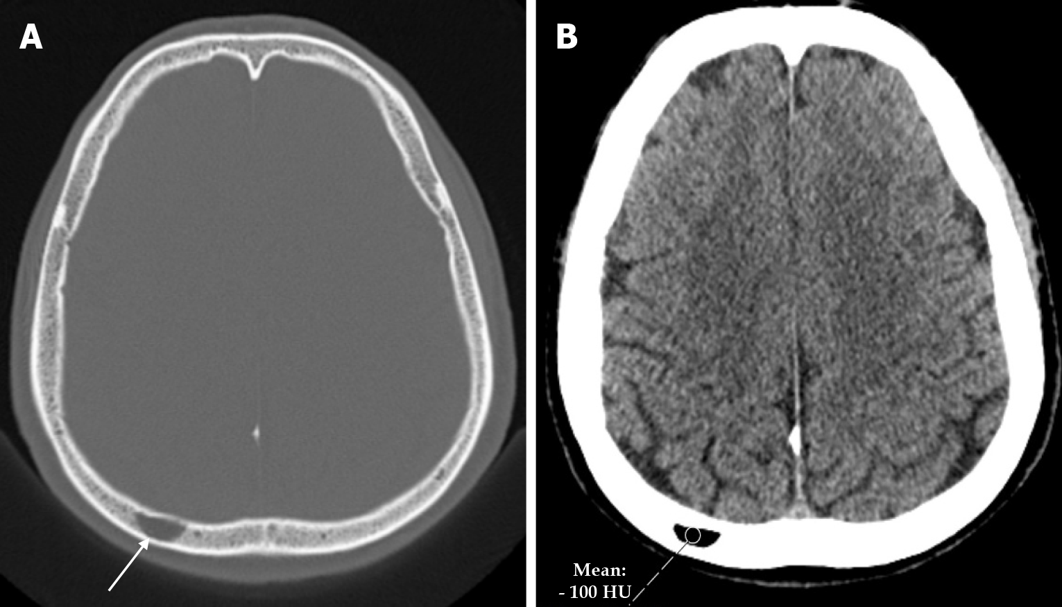
Figure 23 Intraosseous lipoma in fifty-three years old male patient who underwent imaging for headache.
A: Axial computed tomography on the bone window shows a well-circumscribed lesion (arrow) in the diploe of the right parietal bone; B: Axial computed tomography on the parenchymal window shows a lipoma with a mean density of -100 hounsfield unit in a well-circumscribed lesion in the diploe of the right parietal bone.
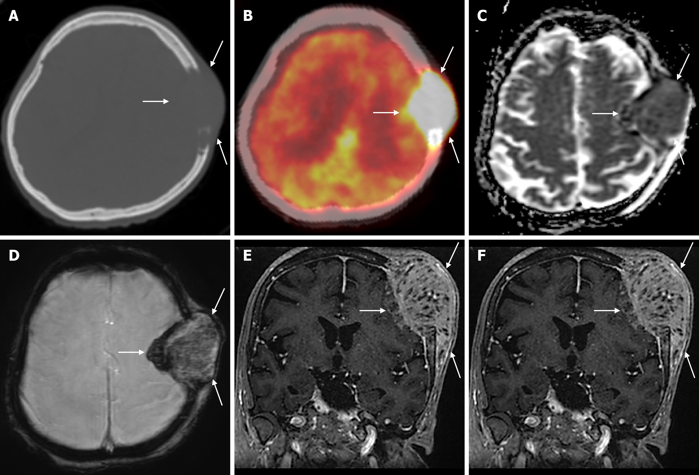
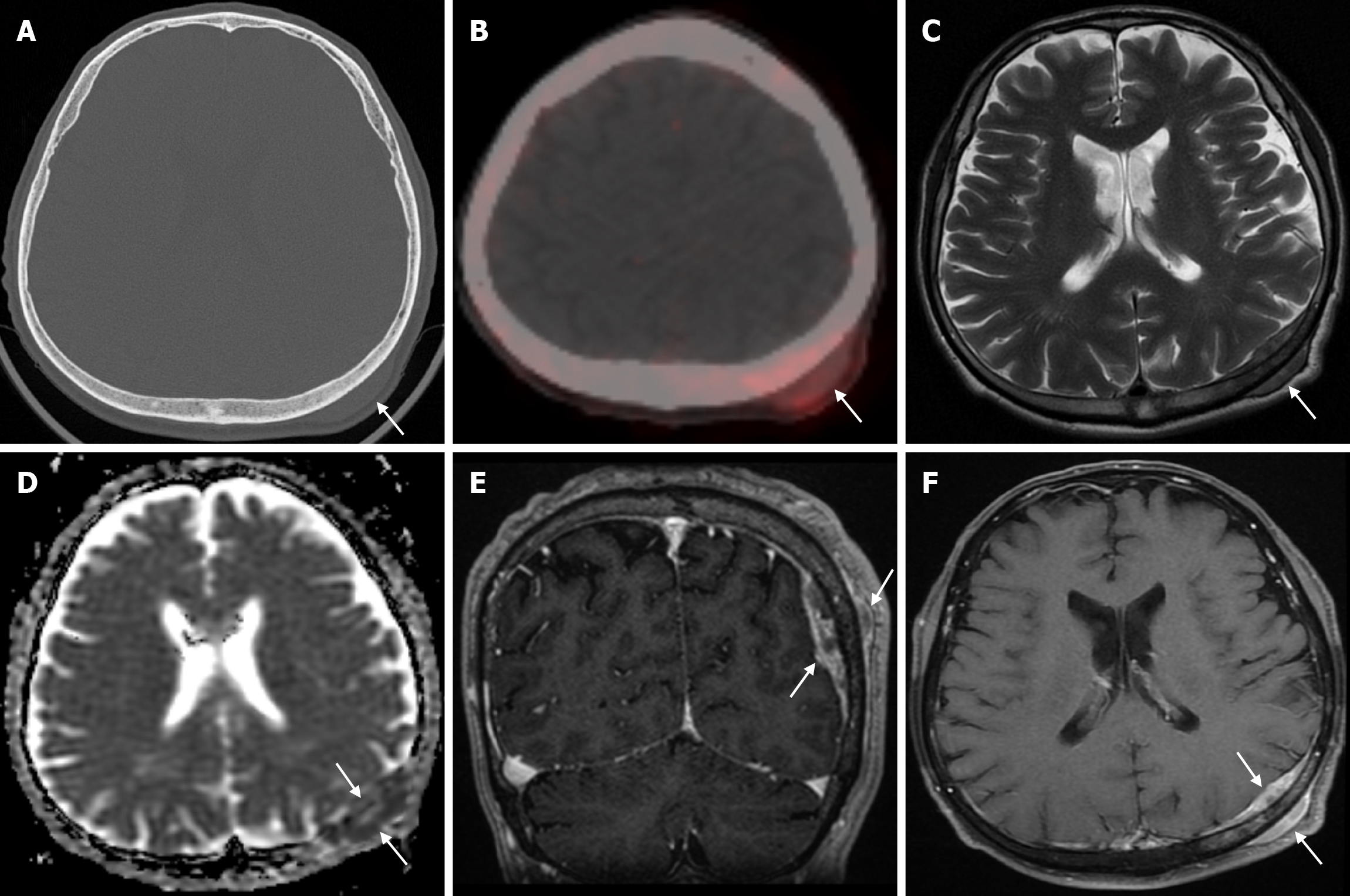
Figure 24 Calvarial solitary lytic metastasis of endometrial carcinoma (Malignant mixed mullerian tumor) in sixty-two years old female patient.
A: Axial computed tomography on the bone window shows a lytic mass lesion (arrows) extending to the scalp, destroying the inner and outer table of the left frontoparietal bone; B: Axial 18F-fluorodeoxyglucose positron emission tomography (18F-FDG PET-CT) shows intense 18F-FDG uptake (SUVmax: 30.13) in the left frontoparietal mass lesion (arrows); C: Apparent diffusion coefficient map shows mild diffusion restriction in the mass; D: Hypointense hemosiderin deposits secondary to bleeding or calcifications products in the mass shows on axial enhanced susceptibility weighted angiography images; E: Coronal contrast enhanced BRAVO image shows heterogeneous, occasionally linear contrast uptake in the mass with periosteal reaction infiltrating the cerebral parenchyma; F: Axial fat-saturated contrast enhanced T1-weighted image shows intense heterogeneous contrast uptake in the metastatic mass (arrows).
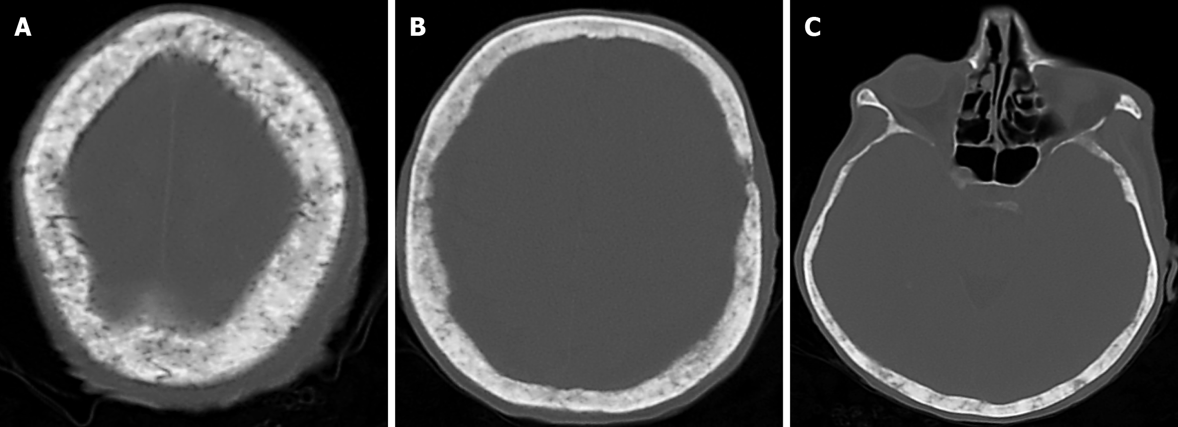
Figure 25 Calvarial diffuse sclerotic metastases of prostate cancer in forty-nine years old male patient.
A-C: Axial computed tomography on the bone window show calvarial diffuse sclerotic metastases.
Figure 26 Calvarial solitary metastasis of a lung neuroendocrine tumor in fifty-nine years old male patient.
A: Axial computed tomography on the bone window shows a soft tissue lesion (arrow) in the extra-axial region between the scalp and the parietal bone on the left parietal side and adjacent to the inner table, without obvious deformation of the bone structure; B: Axial 68Ga-DOTATATE positron emission tomography shows heterogeneous, slightly enhanced uptake (SUVmax: 2.22) in the mass lesion in the left parietal scalp (arrow); C: Axial T2-weighted image shows heterogeneous mildly hypointense signal uptake in the left parietal bone and soft tissue components in the scalp (arrows) with extra-axial distance without significant destruction of the cortical surfaces; D: Diffusion restriction in the mass shows on apparent diffusion coefficient map (arrows); E: Coronal contrast enhanced BRAVO image shows contrast enhancement (arrows) in the parietal bone and soft tissue components; F: Axial fat-saturated contrast enhanced T1-weighted image shows intense heterogeneous contrast enhancement (arrows) in the metastatic mass.
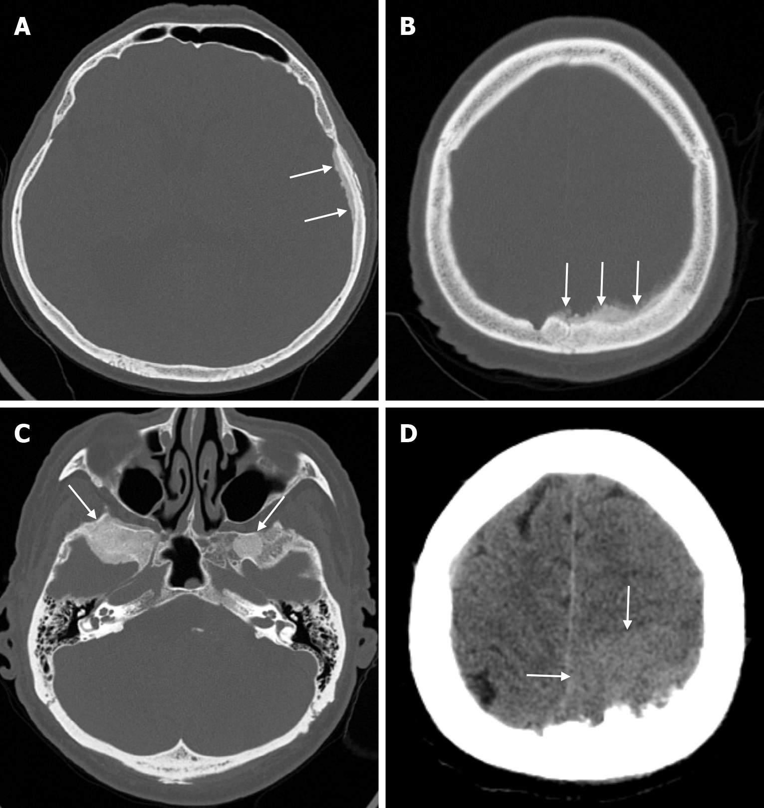
Figure 27 Multifocal sclerotic metastases of prostate cancer in sixty-two years old male patient.
A: Axial computed tomography on the bone window shows metastatic involvement of the left parietal bone causing hyperostosis of the dural surfaces (arrows); B: Axial computed tomography shows bone and dura involvement (arrows) with sclerosis and radial periosteal reaction in the left parietal bone; C: Axial computed tomography on the bone window shows sclerotic metastatic lesions (arrows) in the greater wings of the bilateral sphenoid bones; D: Axial computed tomography on the parenchymal window shows a mass lesion (arrows) with bone and dura involvement and indentation of the parenchyma.
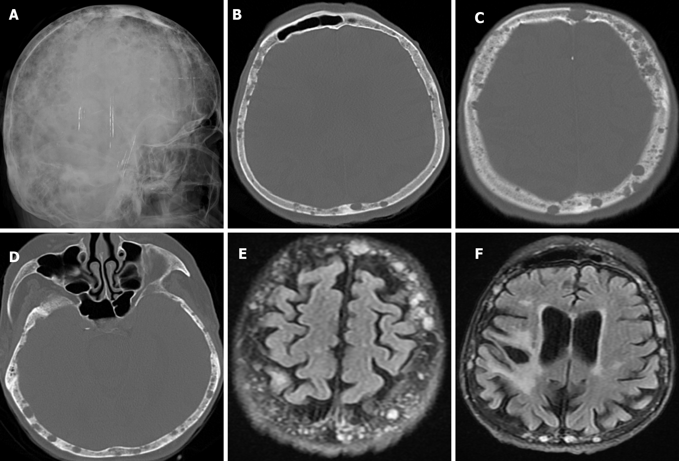
Figure 28 Calvarial involvement of multiple myeloma in ninety-five years old female patient.
A: Lateral plain head radiography shows multiple, small, round, punched-out raindrop-shaped, and sharply circumscribed lesions in the calvarium; B-D: Axial computed tomography on the bone window show multiple, lytic, punched-out, and sharply circumscribed lesions in the calvarial bony structures; E and F: Axial T2 FLAIR images show nodular hyperintense multiple myeloma lesions of different sizes in the calvarium. FLAIR: Fluid-attenuated inversion recovery.
Figure 29 Calvarial involvement of non-Hodgkin's lymphoma (B-cell lymphoma) in fifty-six years old female patient.
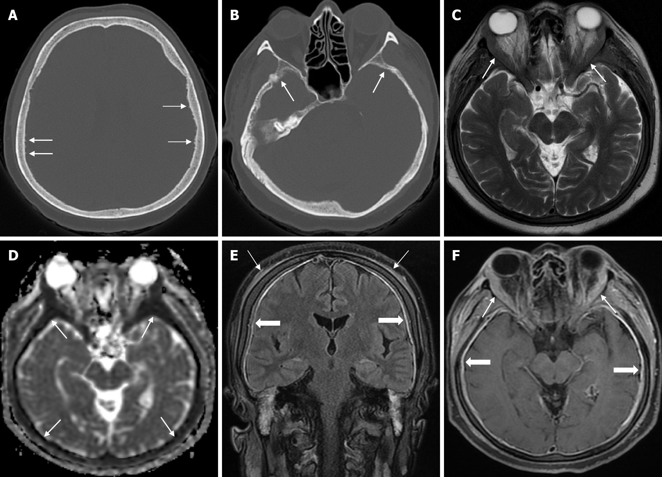
A and B: Axial computed tomography on the bone window show cortical irregularities (thin arrows) due to spiculated periosteal reactions in the inner table; C: Axial T2-weighted image shows symmetrical soft tissue and hypointense signals (thin arrows) in the vicinity of the orbital bones at the superolateral level of both orbits; D: On apparent diffusion coefficient map shows diffuse diffusion restriction at the diploe distance (thin arrows); E: Coronal contrast enhanced fluid-attenuated inversion recovery T2-weighted image shows diffuse periosteal thickening (solid periosteal reactions) and periosteal contrast enhancement (thin arrows) that may be secondary to inflammatory hyperemia and diffuse dural contrast enhancement (thick arrows); F: Axial fat-saturated contrast enhanced T1-weighted image shows lesions with contrast enhancement and diffuse dural (nodular and smooth) contrast enhancement (thick arrows) with continuity along the inner and outer table in the cranial bones, accompanied by soft tissue components surrounding the bones (thin arrows).
Figure 30 Neuroblastoma metastasis in two years old female patient.
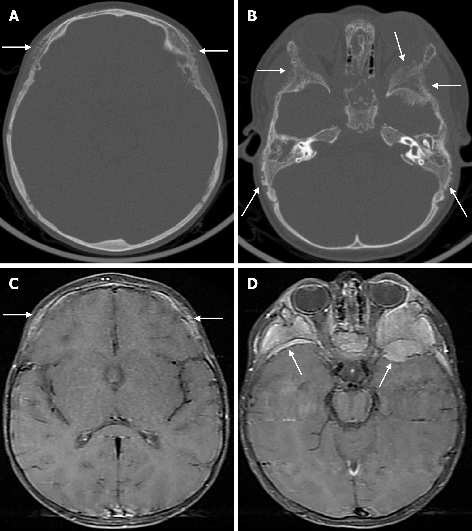
A and B: Axial computed tomography on the bone window show spiculated periosteal reactions (arrows) in the calvarial table, more prominent in the outer table, secondary to metastatic involvement of bilateral frontal, parietal, temporal and sphenoid bones; C and D: Axial fat-saturated contrast enhanced T1-weighted image show mass lesions (arrows) consistent with tumoral metastatic infiltration with intense contrast enhancement accompanied by soft tissue components in bilateral frontal, parietal, temporal and sphenoid bones.
- Citation: Gökçe E, Beyhan M. Review of imaging modalities and radiological findings of calvarial lesions. World J Radiol 2025; 17(6): 107776
- URL: https://www.wjgnet.com/1949-8470/full/v17/i6/107776.htm
- DOI: https://dx.doi.org/10.4329/wjr.v17.i6.107776


