Copyright
©The Author(s) 2025.
World J Radiol. Jun 28, 2025; 17(6): 107776
Published online Jun 28, 2025. doi: 10.4329/wjr.v17.i6.107776
Published online Jun 28, 2025. doi: 10.4329/wjr.v17.i6.107776
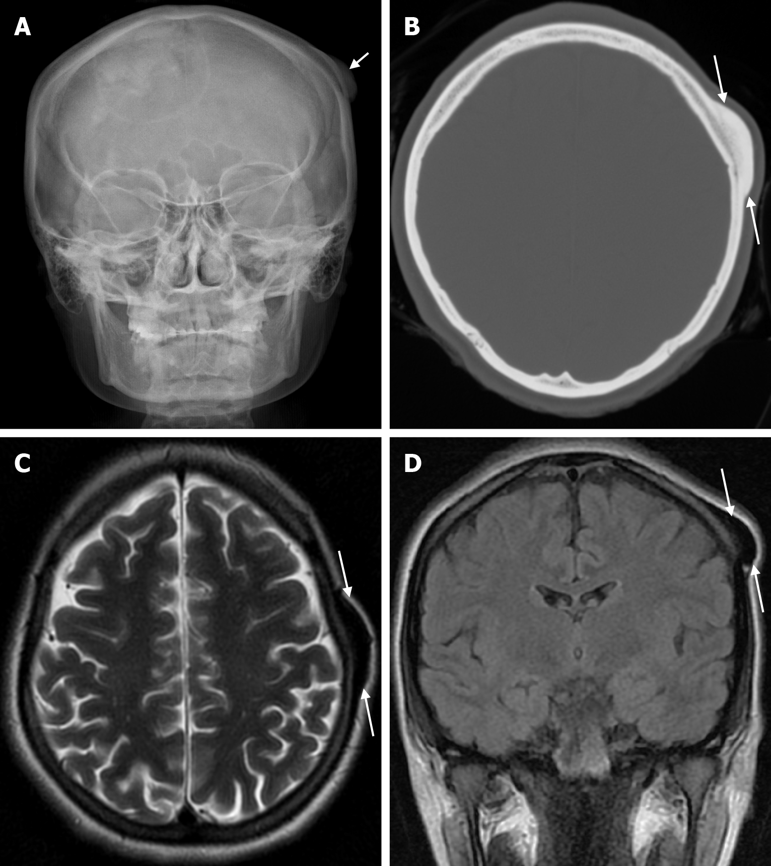
Figure 17 Osteoma in twenty-five years old female patient who underwent imaging for swelling of the scalp.
A: Plain anteroposterior head radiography shows a smooth contoured lesion (arrow) in a compact bony structure extending from the calvarial outer table; B: Axial computed tomography on the bone window shows an isodense smooth contoured lesion (arrows) with broad-based bony structures extending from the outer table to the scalp in the parietal bone; C and D: The left parietal bone shows a smooth contoured sclerotic lesion (arrows) with protrusion from the outer table to the scalp, with a prominent hypointense signal on T2-weighted images.
- Citation: Gökçe E, Beyhan M. Review of imaging modalities and radiological findings of calvarial lesions. World J Radiol 2025; 17(6): 107776
- URL: https://www.wjgnet.com/1949-8470/full/v17/i6/107776.htm
- DOI: https://dx.doi.org/10.4329/wjr.v17.i6.107776


