Copyright
©The Author(s) 2025.
World J Radiol. Jun 28, 2025; 17(6): 107776
Published online Jun 28, 2025. doi: 10.4329/wjr.v17.i6.107776
Published online Jun 28, 2025. doi: 10.4329/wjr.v17.i6.107776
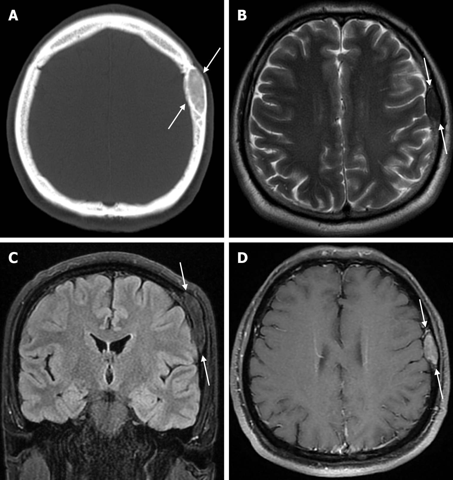
Figure 18 Fibrous dysplasia in thirty-five years old male patient who underwent imaging for headache.
A: Axial computed tomography on the bone window shows a smooth contoured lesion with ground glass density (arrows) in the left parietal bone; B: On axial T2-weighted image, hypointense lesion in the parietal bone (arrows); C: On coronal FLAIR T2-weighted image, mild hyperintense lesion in the parietal bone (arrows); D: Axial contrast enhanced T1-weighted image shows a well-circumscribed lesion (arrows) with contrast enhancement in the parietal bone. FLAIR: Fluid-attenuated inversion recovery.
- Citation: Gökçe E, Beyhan M. Review of imaging modalities and radiological findings of calvarial lesions. World J Radiol 2025; 17(6): 107776
- URL: https://www.wjgnet.com/1949-8470/full/v17/i6/107776.htm
- DOI: https://dx.doi.org/10.4329/wjr.v17.i6.107776


