Copyright
©The Author(s) 2025.
World J Radiol. Jun 28, 2025; 17(6): 107776
Published online Jun 28, 2025. doi: 10.4329/wjr.v17.i6.107776
Published online Jun 28, 2025. doi: 10.4329/wjr.v17.i6.107776
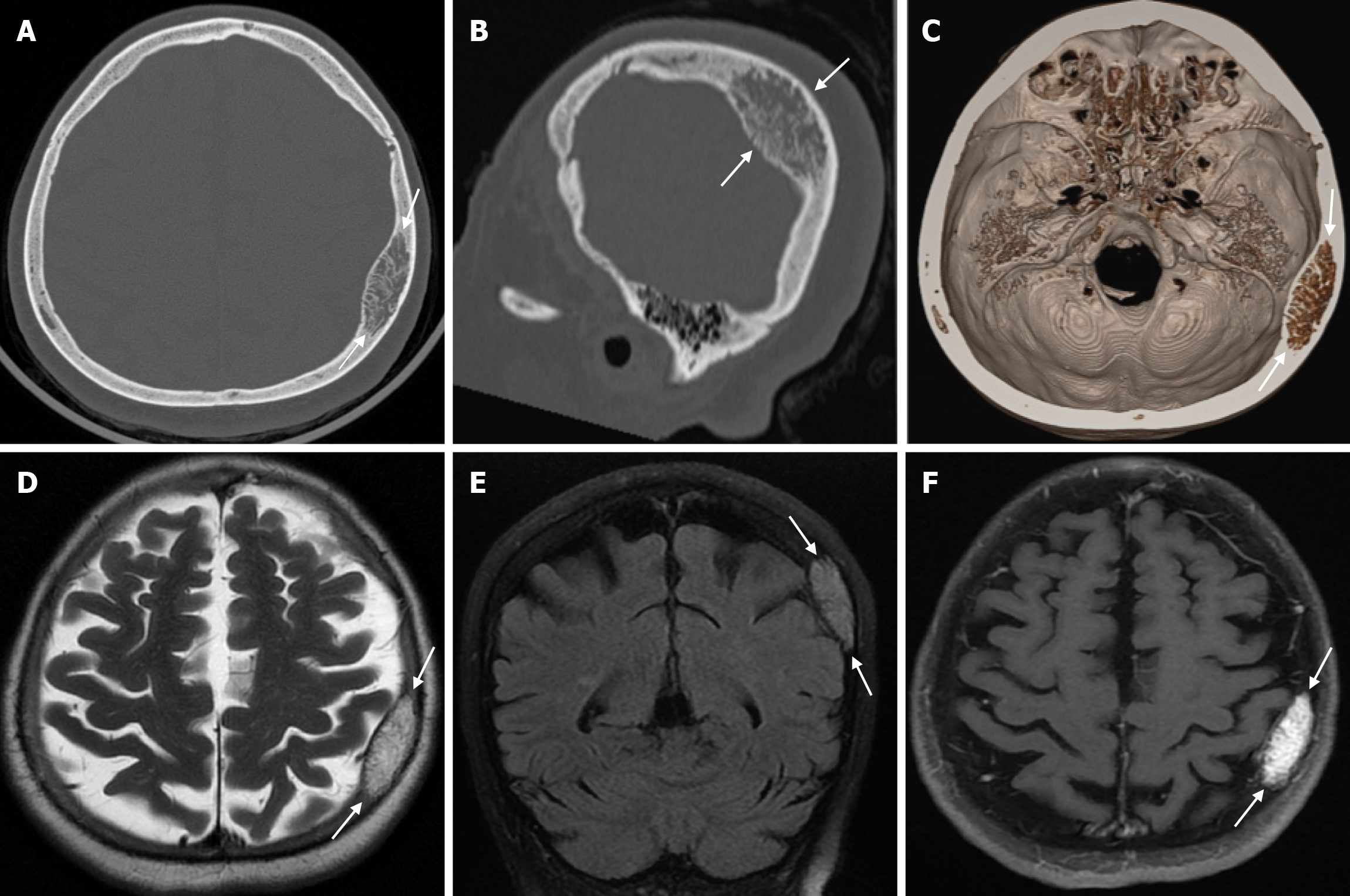
Figure 22 Intraosseous venous malformation in seventy-one years old female patient who underwent imaging for headache.
A and B: Sagittal computed tomography on the bone window show a lytic lesion (arrows) in the left parietal bone, localized in the diploe region, causing widening of the inner table, in which bone trabeculae can be intensely selected; C: Three-dimensional computed tomography shows an intraosseous venous malformation (arrows) in the left parietal bone; D: The lesion is hyperintense (arrows) on axial T2-weighted image; E: On coronal fluid-attenuated inversion recovery T2-weighted image the lesion is hyperintense (arrows); F: On axial contrast enhanced T1-weighted image the lesion shows intense contrast enhancement (arrows).
- Citation: Gökçe E, Beyhan M. Review of imaging modalities and radiological findings of calvarial lesions. World J Radiol 2025; 17(6): 107776
- URL: https://www.wjgnet.com/1949-8470/full/v17/i6/107776.htm
- DOI: https://dx.doi.org/10.4329/wjr.v17.i6.107776


