Copyright
©The Author(s) 2025.
World J Radiol. Jun 28, 2025; 17(6): 107776
Published online Jun 28, 2025. doi: 10.4329/wjr.v17.i6.107776
Published online Jun 28, 2025. doi: 10.4329/wjr.v17.i6.107776
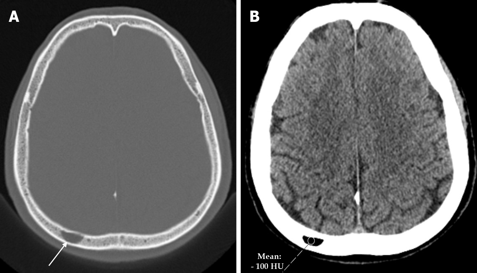
Figure 23 Intraosseous lipoma in fifty-three years old male patient who underwent imaging for headache.
A: Axial computed tomography on the bone window shows a well-circumscribed lesion (arrow) in the diploe of the right parietal bone; B: Axial computed tomography on the parenchymal window shows a lipoma with a mean density of -100 hounsfield unit in a well-circumscribed lesion in the diploe of the right parietal bone.
- Citation: Gökçe E, Beyhan M. Review of imaging modalities and radiological findings of calvarial lesions. World J Radiol 2025; 17(6): 107776
- URL: https://www.wjgnet.com/1949-8470/full/v17/i6/107776.htm
- DOI: https://dx.doi.org/10.4329/wjr.v17.i6.107776


