Copyright
©The Author(s) 2025.
World J Radiol. Jun 28, 2025; 17(6): 107776
Published online Jun 28, 2025. doi: 10.4329/wjr.v17.i6.107776
Published online Jun 28, 2025. doi: 10.4329/wjr.v17.i6.107776
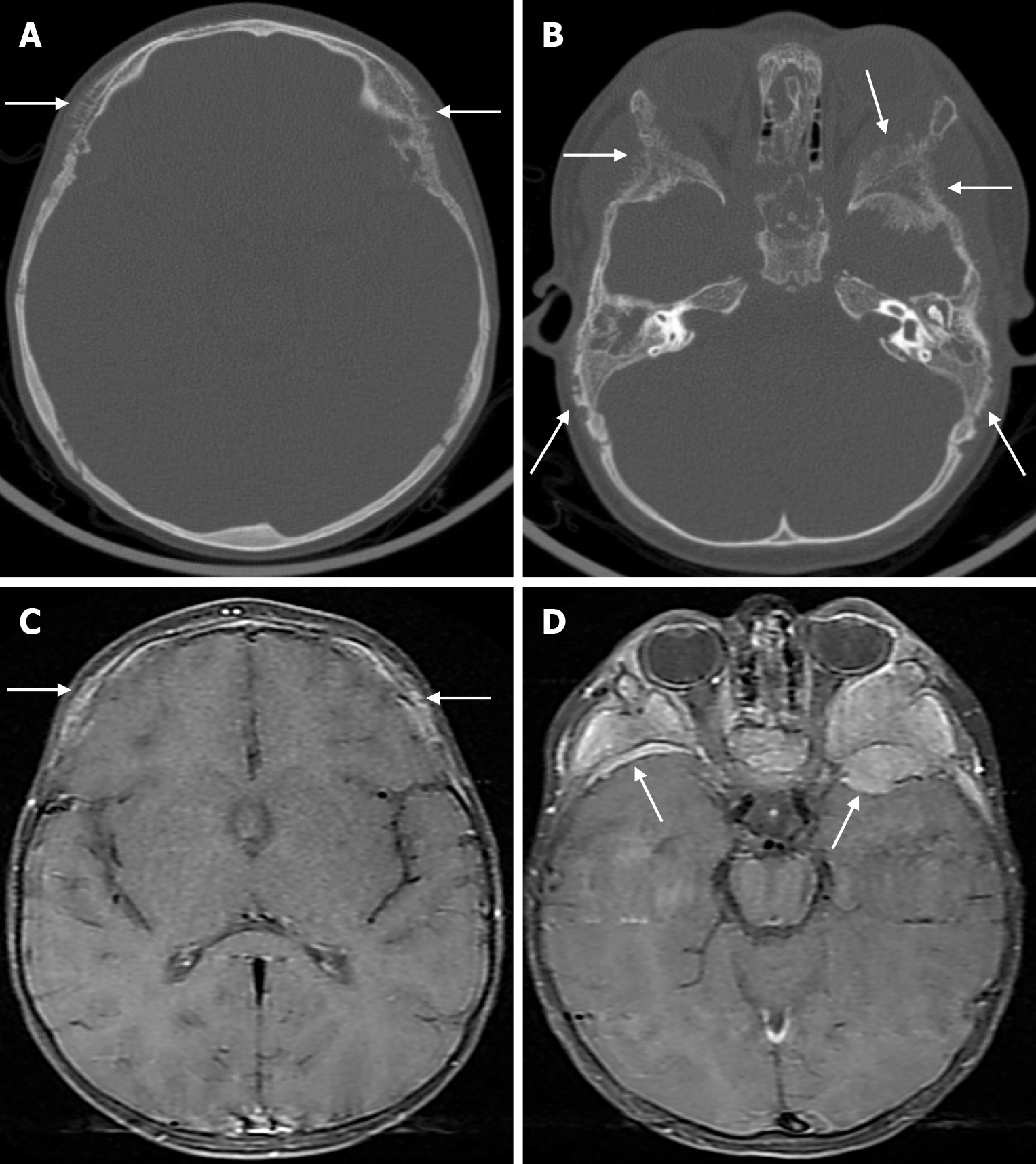
Figure 30 Neuroblastoma metastasis in two years old female patient.
A and B: Axial computed tomography on the bone window show spiculated periosteal reactions (arrows) in the calvarial table, more prominent in the outer table, secondary to metastatic involvement of bilateral frontal, parietal, temporal and sphenoid bones; C and D: Axial fat-saturated contrast enhanced T1-weighted image show mass lesions (arrows) consistent with tumoral metastatic infiltration with intense contrast enhancement accompanied by soft tissue components in bilateral frontal, parietal, temporal and sphenoid bones.
- Citation: Gökçe E, Beyhan M. Review of imaging modalities and radiological findings of calvarial lesions. World J Radiol 2025; 17(6): 107776
- URL: https://www.wjgnet.com/1949-8470/full/v17/i6/107776.htm
- DOI: https://dx.doi.org/10.4329/wjr.v17.i6.107776


