Copyright
©The Author(s) 2025.
World J Radiol. Jun 28, 2025; 17(6): 107776
Published online Jun 28, 2025. doi: 10.4329/wjr.v17.i6.107776
Published online Jun 28, 2025. doi: 10.4329/wjr.v17.i6.107776
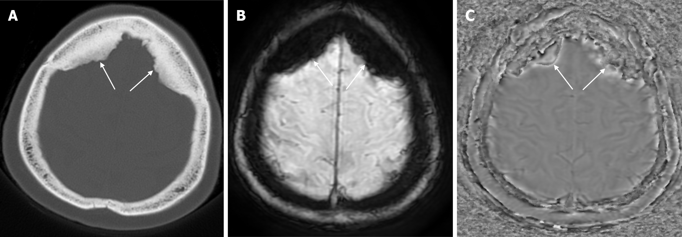
Figure 11 Hyperostosis frontalis interna in sixty-seven years old female patient who underwent imaging for headache.
A: Axial computed tomography on the bone window shows symmetrical calvarial thickening (arrows) of similar density to the bone extending from the inner table to the extra-axial space in the bilateral frontal bones; B: Hyperostosis frontalis interna is hypointense on axial enhanced susceptibility weighted angiography (eSWAN) images, C: Hyperintense on axial filtered phase eSWAN images, signal intensities compatible with hyperosteosis frontalis interna (arrows) at the level of the inner table of the frontal bone.
- Citation: Gökçe E, Beyhan M. Review of imaging modalities and radiological findings of calvarial lesions. World J Radiol 2025; 17(6): 107776
- URL: https://www.wjgnet.com/1949-8470/full/v17/i6/107776.htm
- DOI: https://dx.doi.org/10.4329/wjr.v17.i6.107776


