Copyright
©The Author(s) 2025.
World J Radiol. Jun 28, 2025; 17(6): 107776
Published online Jun 28, 2025. doi: 10.4329/wjr.v17.i6.107776
Published online Jun 28, 2025. doi: 10.4329/wjr.v17.i6.107776
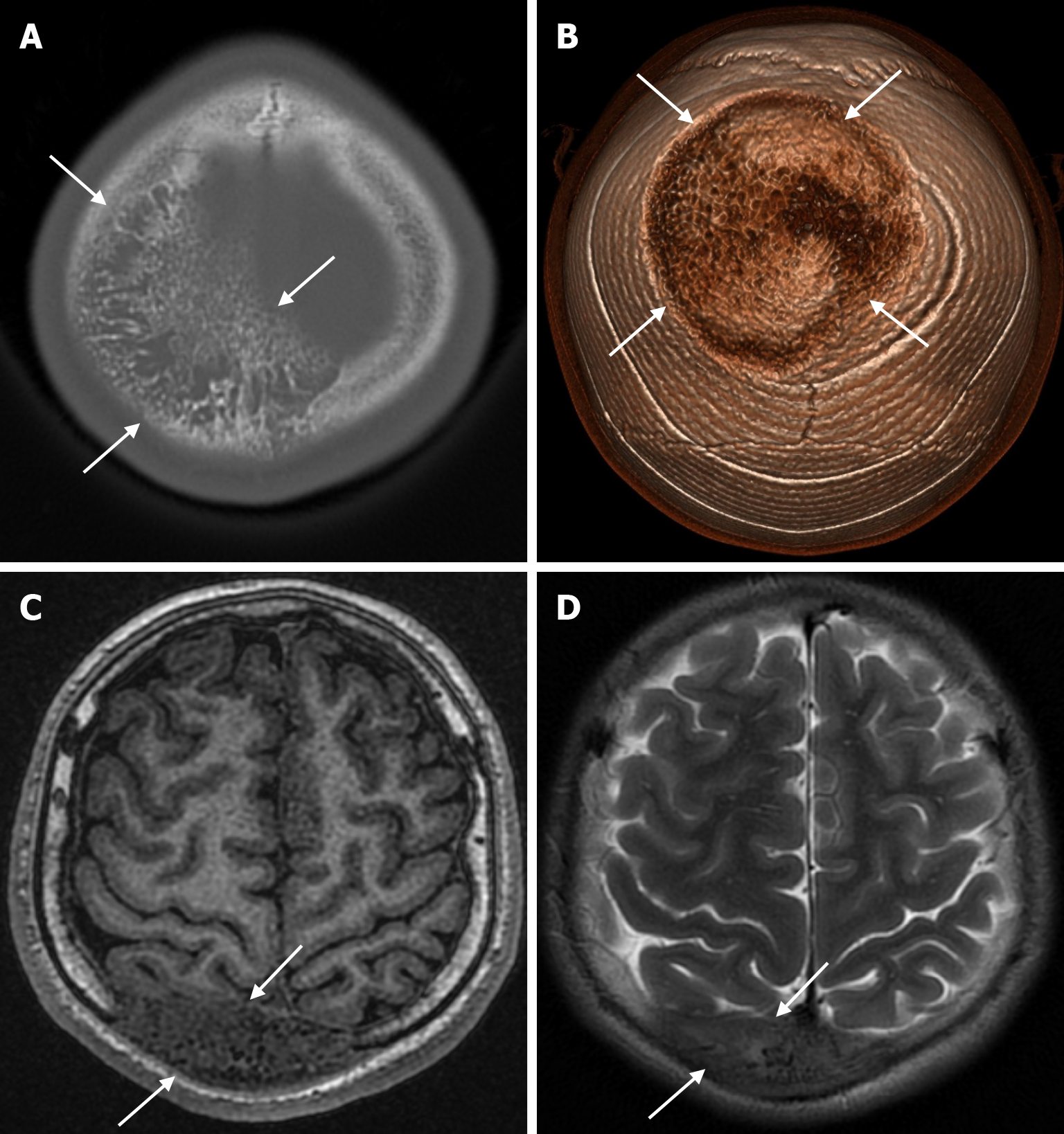
Figure 20 Fibrous dysplasia in seventeen years old male patient who underwent imaging for a calvarial mass.
A: Axial computed tomography on the bone window shows fibrous dysplasia (arrows) mimicking intraosseous venous malformation in both parietal bones, predominantly involving the right parietal bone in the plane of convexity, with coarse trabecular bone structures and lytic areas in a radial pattern involving the inner and outer table and protruding beyond the bone; B: Three-dimensional computed tomography shows fibrous dysplasia protruding from both parietal bones (arrows); C: On axial T1-weighted image, the lesion is heterogeneously hypointense in both parietal bones (arrows); D: Axial T2-weighted image shows heterogeneous mild hyperintensity (arrows) in both parietal bones.
- Citation: Gökçe E, Beyhan M. Review of imaging modalities and radiological findings of calvarial lesions. World J Radiol 2025; 17(6): 107776
- URL: https://www.wjgnet.com/1949-8470/full/v17/i6/107776.htm
- DOI: https://dx.doi.org/10.4329/wjr.v17.i6.107776


