Copyright
©The Author(s) 2025.
World J Radiol. Jun 28, 2025; 17(6): 107776
Published online Jun 28, 2025. doi: 10.4329/wjr.v17.i6.107776
Published online Jun 28, 2025. doi: 10.4329/wjr.v17.i6.107776
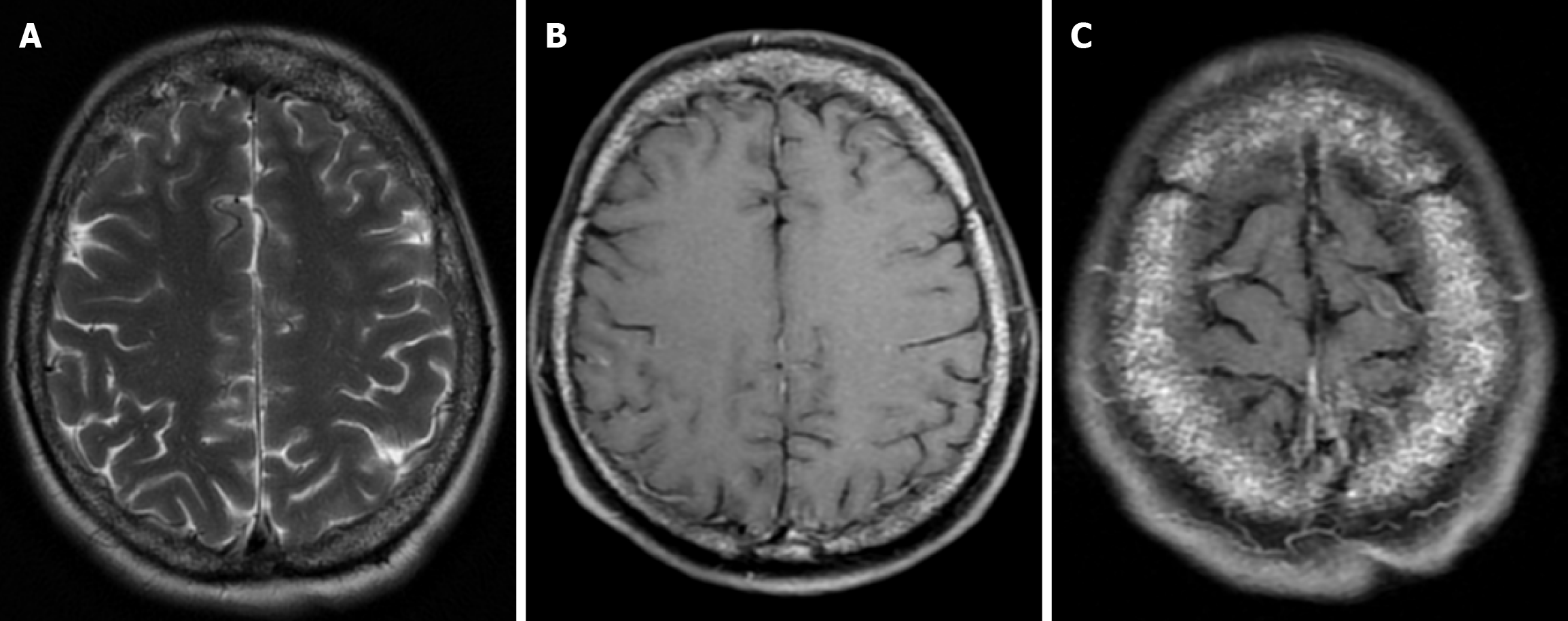
Figure 14 Thalassemia major with calvarial involvement in forty-three years old female patient.
A: On axial T2-weighted image, the skull bones show heterogeneous signal intensity at diploe distance; B and C: On axial fat-saturated contrast enhanced T1-weighted image, the skull bones show bone marrow lesions secondary to thalassaemia major with diffuse heterogeneous contrast enhancement.
- Citation: Gökçe E, Beyhan M. Review of imaging modalities and radiological findings of calvarial lesions. World J Radiol 2025; 17(6): 107776
- URL: https://www.wjgnet.com/1949-8470/full/v17/i6/107776.htm
- DOI: https://dx.doi.org/10.4329/wjr.v17.i6.107776


