Copyright
©The Author(s) 2025.
World J Radiol. Jun 28, 2025; 17(6): 107776
Published online Jun 28, 2025. doi: 10.4329/wjr.v17.i6.107776
Published online Jun 28, 2025. doi: 10.4329/wjr.v17.i6.107776
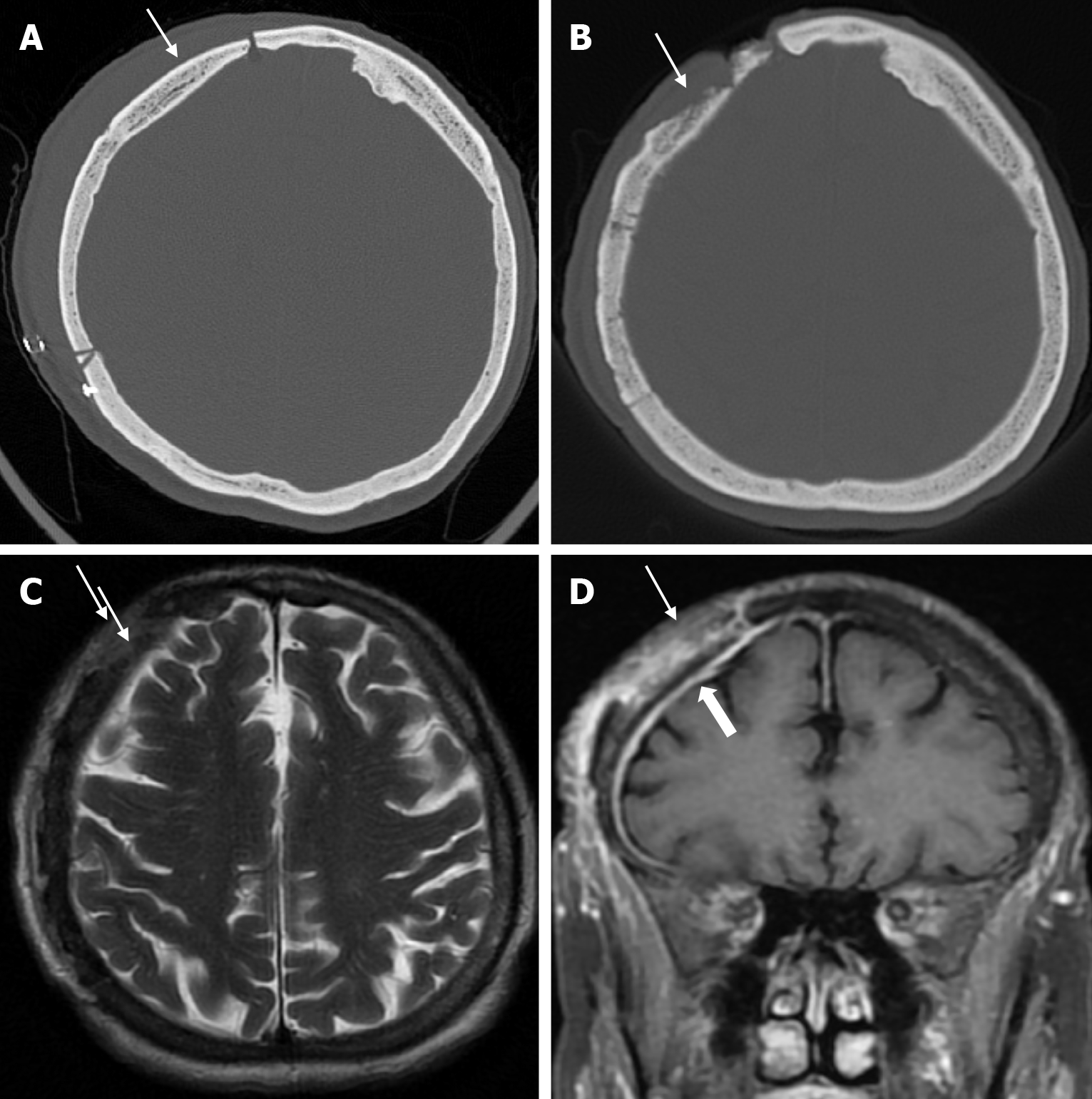
Figure 12 Calvarial osteomyelitis in sixty-nine years old female who underwent surgery for a subdural hematoma.
A: Axial computed tomography of the bone window shows a defect in the right frontoparietal bone secondary to the previous surgery and the outer calvarial table (thin arrow) is intact; B: Axial computed tomography of the bone window six months later shows irregularity and thinning of the outer table (thin arrow) at the surgical site; C: Axial T2-weighted image shows hyperintensity (thin arrow) in the frontoparietal bone; D: On coronal contrast enhanced T1-weighted image, irregularities in the frontoparietal cortical surfaces and heterogeneous contrast enhancement in the bone (thin arrow) and contrast enhancement in the adjacent dura (thick arrow).
- Citation: Gökçe E, Beyhan M. Review of imaging modalities and radiological findings of calvarial lesions. World J Radiol 2025; 17(6): 107776
- URL: https://www.wjgnet.com/1949-8470/full/v17/i6/107776.htm
- DOI: https://dx.doi.org/10.4329/wjr.v17.i6.107776


