Copyright
©The Author(s) 2025.
World J Radiol. Jun 28, 2025; 17(6): 107776
Published online Jun 28, 2025. doi: 10.4329/wjr.v17.i6.107776
Published online Jun 28, 2025. doi: 10.4329/wjr.v17.i6.107776
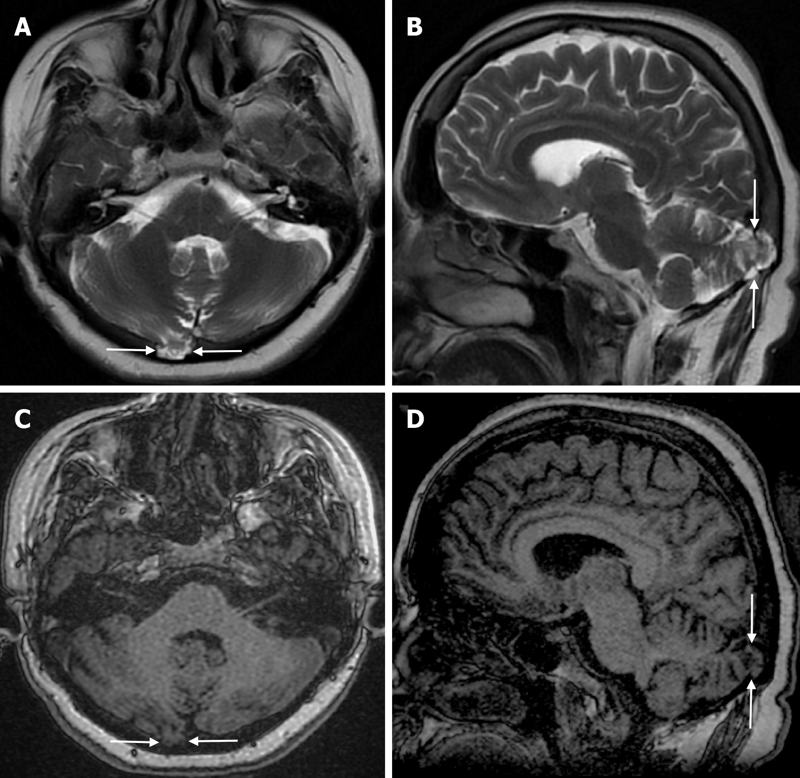
Figure 2 Cerebellar parenchyma herniations into the arachnoid granulations fifty-four-year-old female patient with no history of trauma who underwent imaging for headache.
A: On axial T2-weighted image; B: On sagittal T2-weighted image; C: On axial T1-weighted image; D: On sagittal T1-weighted image, the defect with a maximum diameter of 1.5 centimeter in the inner table to the right of the midline in the right occipital bone and signal intensities of the adjacent cerebellar parenchyma and arachnoid granulation herniating into the diploic space are shown (arrows).
- Citation: Gökçe E, Beyhan M. Review of imaging modalities and radiological findings of calvarial lesions. World J Radiol 2025; 17(6): 107776
- URL: https://www.wjgnet.com/1949-8470/full/v17/i6/107776.htm
- DOI: https://dx.doi.org/10.4329/wjr.v17.i6.107776


