Copyright
©The Author(s) 2025.
World J Radiol. Jun 28, 2025; 17(6): 107776
Published online Jun 28, 2025. doi: 10.4329/wjr.v17.i6.107776
Published online Jun 28, 2025. doi: 10.4329/wjr.v17.i6.107776
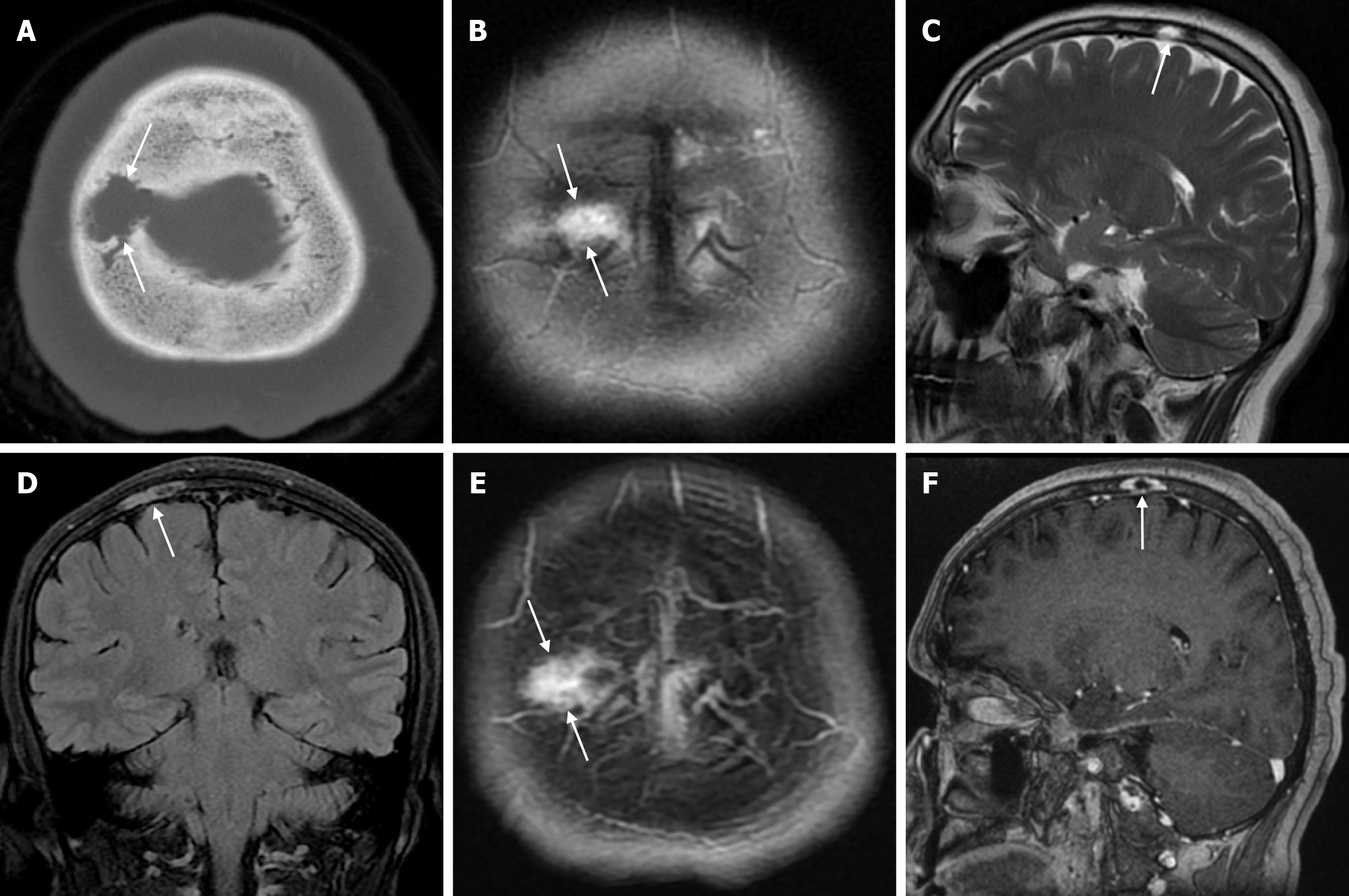
Figure 3 Venous lake in fifty-four years old female patient who underwent imaging for headache.
A; Axial computed tomography on the bone window shows irregular circumscribed structures (arrows) extending to the diploe distance in the right parietal bone in the plane of convexity, without destruction of the outer table; B: On axial T2-weighted image; C: On sagittal T2-weighted image; D: On coronal FLAIR T2-weighted image; E: On axial contrast enhanced T1-weighted image; F: On sagittal contrast enhanced T1-weighted image, in the parasagittal right parietal bone in the plane of convexity, hyperintense on T2-weighted series, hypointense on T1-weighted series, contrast enhanced venous lake (arrows) located in the diploe distance with mild focal irregularity in the inner table. FLAIR: Fluid-attenuated inversion recovery.
- Citation: Gökçe E, Beyhan M. Review of imaging modalities and radiological findings of calvarial lesions. World J Radiol 2025; 17(6): 107776
- URL: https://www.wjgnet.com/1949-8470/full/v17/i6/107776.htm
- DOI: https://dx.doi.org/10.4329/wjr.v17.i6.107776


