Copyright
©The Author(s) 2025.
World J Radiol. Jun 28, 2025; 17(6): 107776
Published online Jun 28, 2025. doi: 10.4329/wjr.v17.i6.107776
Published online Jun 28, 2025. doi: 10.4329/wjr.v17.i6.107776
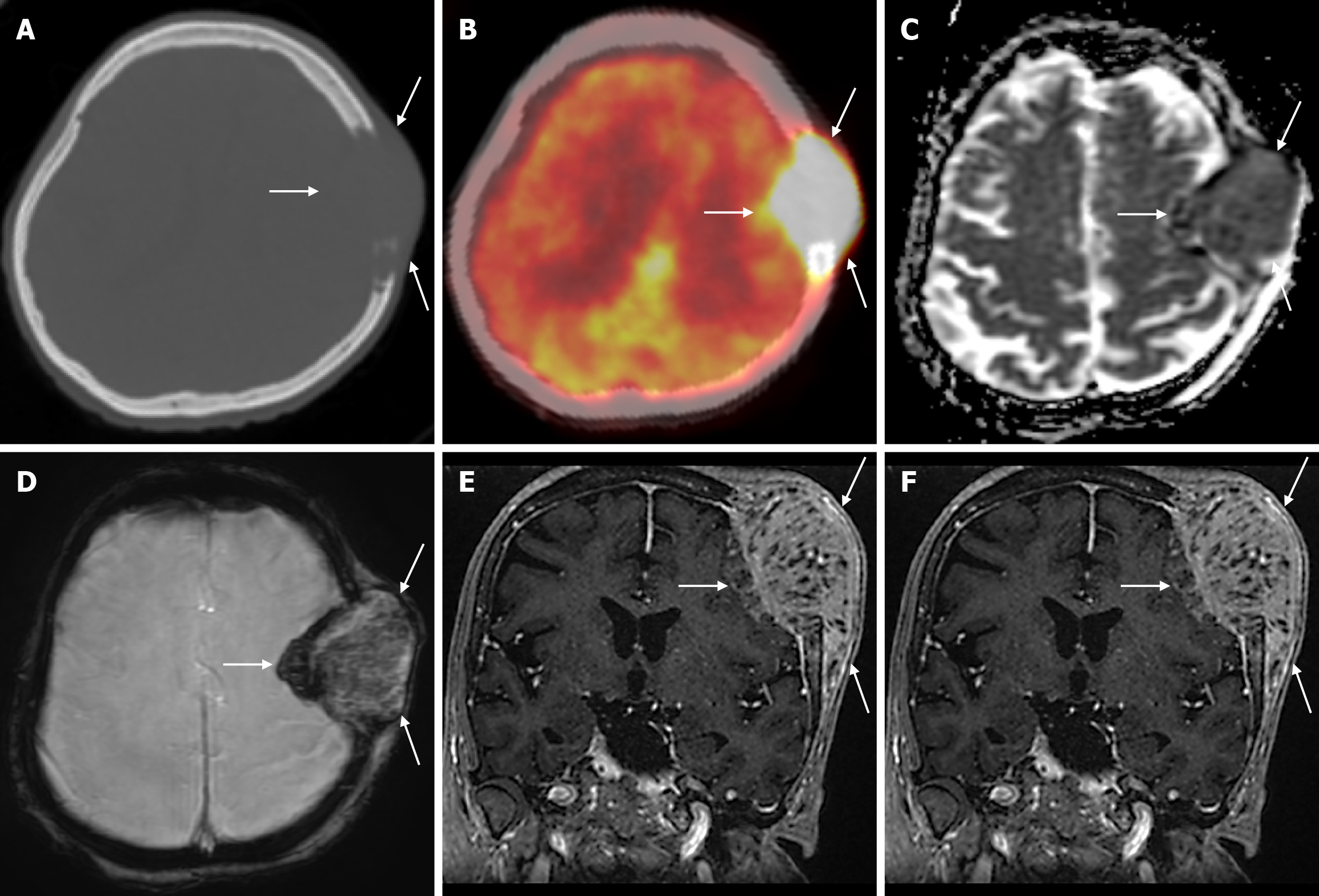
Figure 24 Calvarial solitary lytic metastasis of endometrial carcinoma (Malignant mixed mullerian tumor) in sixty-two years old female patient.
A: Axial computed tomography on the bone window shows a lytic mass lesion (arrows) extending to the scalp, destroying the inner and outer table of the left frontoparietal bone; B: Axial 18F-fluorodeoxyglucose positron emission tomography (18F-FDG PET-CT) shows intense 18F-FDG uptake (SUVmax: 30.13) in the left frontoparietal mass lesion (arrows); C: Apparent diffusion coefficient map shows mild diffusion restriction in the mass; D: Hypointense hemosiderin deposits secondary to bleeding or calcifications products in the mass shows on axial enhanced susceptibility weighted angiography images; E: Coronal contrast enhanced BRAVO image shows heterogeneous, occasionally linear contrast uptake in the mass with periosteal reaction infiltrating the cerebral parenchyma; F: Axial fat-saturated contrast enhanced T1-weighted image shows intense heterogeneous contrast uptake in the metastatic mass (arrows).
- Citation: Gökçe E, Beyhan M. Review of imaging modalities and radiological findings of calvarial lesions. World J Radiol 2025; 17(6): 107776
- URL: https://www.wjgnet.com/1949-8470/full/v17/i6/107776.htm
- DOI: https://dx.doi.org/10.4329/wjr.v17.i6.107776


