Copyright
©The Author(s) 2025.
World J Radiol. Jun 28, 2025; 17(6): 107776
Published online Jun 28, 2025. doi: 10.4329/wjr.v17.i6.107776
Published online Jun 28, 2025. doi: 10.4329/wjr.v17.i6.107776
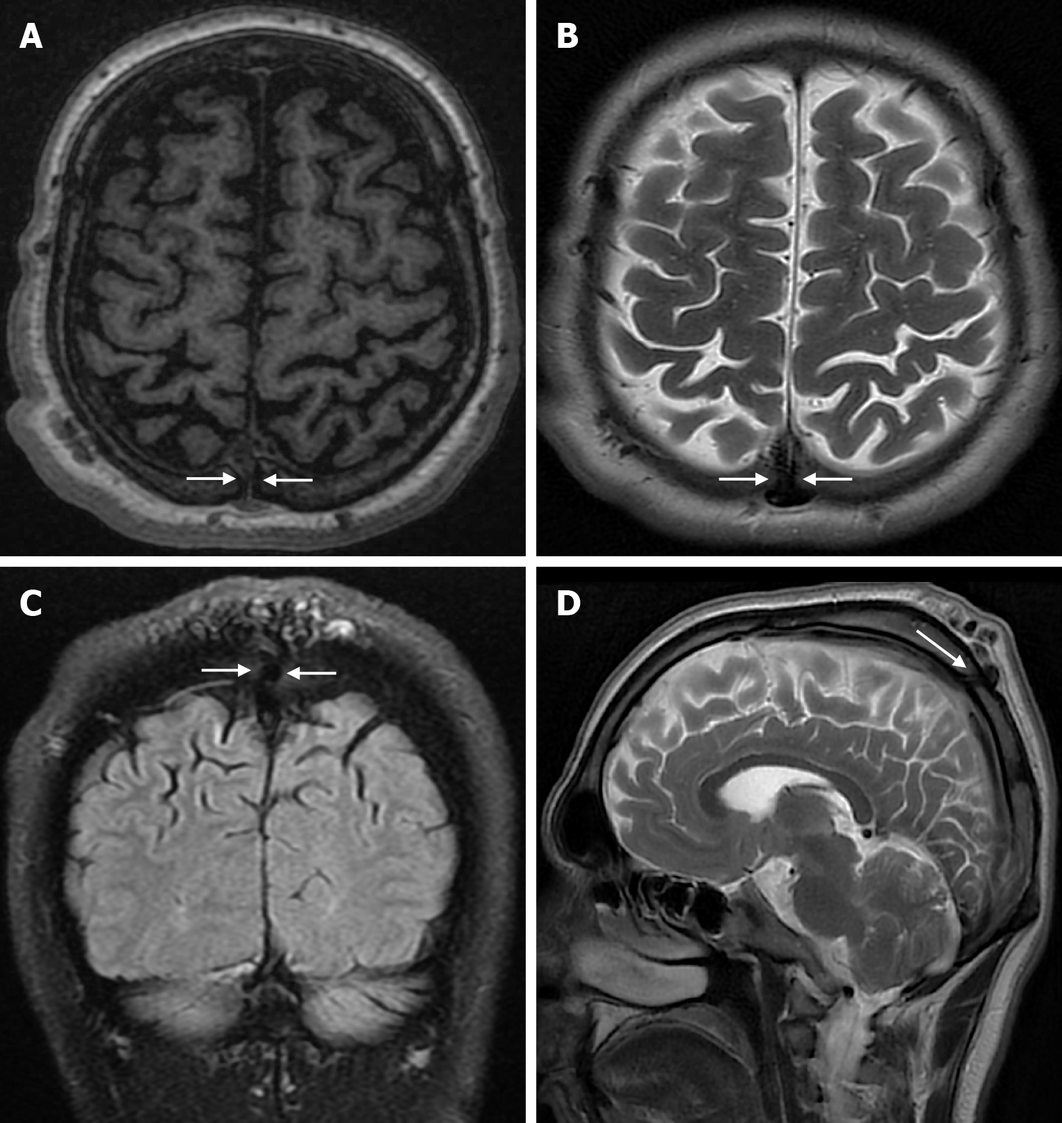
Figure 7 Sinus pericranii in twenty years old male patient who underwent imaging for headache.
A: On axial T1-weighted image; B: On axial T2-weighted image; C: On coronal FLAIR T2-weighted image; D: On sagittal T2-weighted image shows a sinus pericranii associated with the superior sagittal sinus leading to enlarged tortuous venous structures extending to the scalp with a transcarvalial venous duct (arrows) in the midline parietal bone.
- Citation: Gökçe E, Beyhan M. Review of imaging modalities and radiological findings of calvarial lesions. World J Radiol 2025; 17(6): 107776
- URL: https://www.wjgnet.com/1949-8470/full/v17/i6/107776.htm
- DOI: https://dx.doi.org/10.4329/wjr.v17.i6.107776


