Copyright
©The Author(s) 2025.
World J Radiol. Jun 28, 2025; 17(6): 107776
Published online Jun 28, 2025. doi: 10.4329/wjr.v17.i6.107776
Published online Jun 28, 2025. doi: 10.4329/wjr.v17.i6.107776
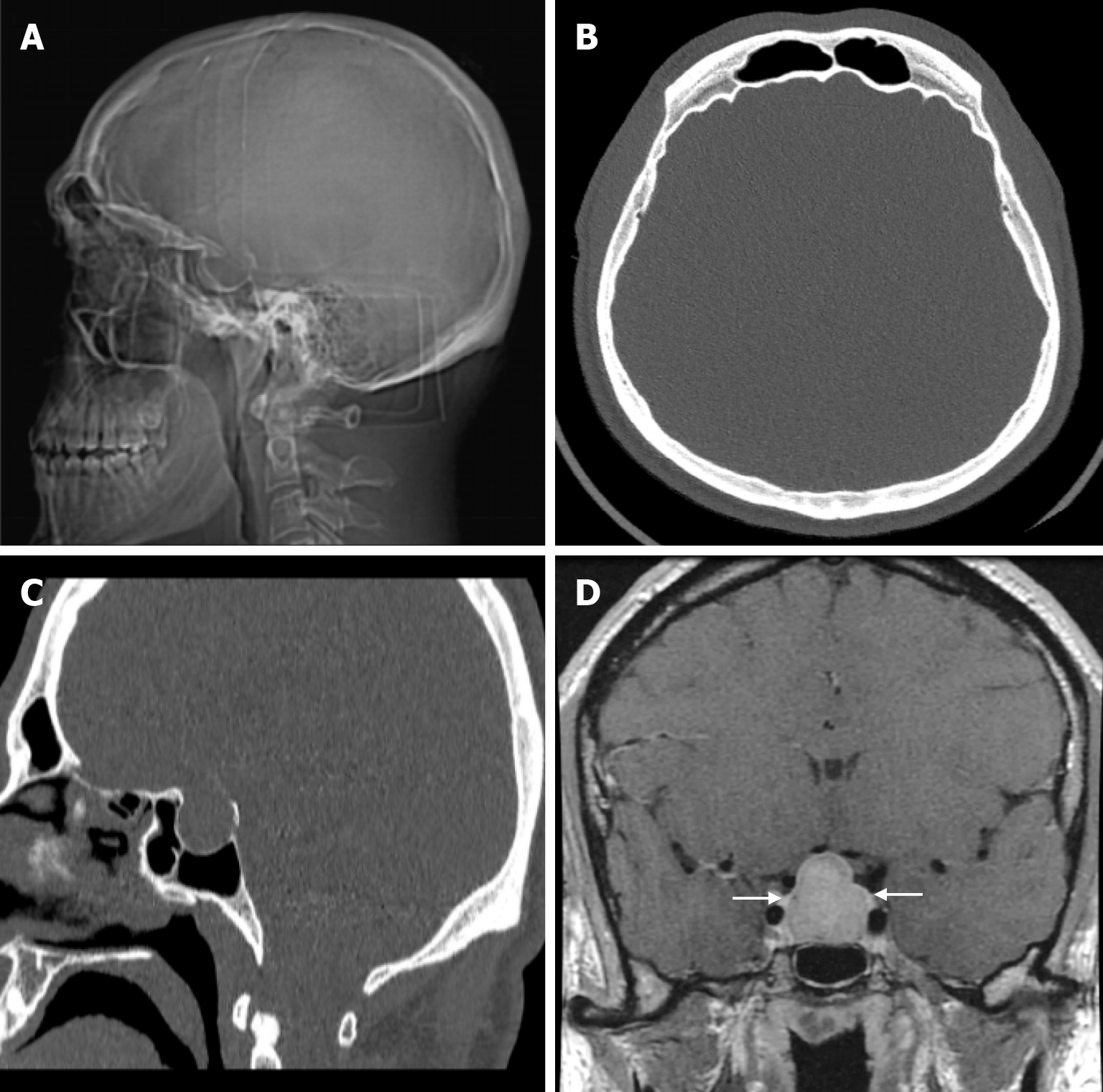
Figure 16 Acromegaly in thirty-two years old male patient who underwent imaging for gigantism.
A: Plain radiography shows thickening of all calvarial bones and expansion of the sella cavity; B: Axial computed tomography on the bone window shows thickening of all calvarial bones; C: Sagittal computed tomography on the bone window shows expansion of the sella cavity; D: Coronal contrast enhanced T1-weighted image shows contrast enhancement macroadenoma (arrows) with smooth lobulated contour completely filling and enlarging the sella cavity, obliterating the suprasellar cistern and compressing the optic chiasm.
- Citation: Gökçe E, Beyhan M. Review of imaging modalities and radiological findings of calvarial lesions. World J Radiol 2025; 17(6): 107776
- URL: https://www.wjgnet.com/1949-8470/full/v17/i6/107776.htm
- DOI: https://dx.doi.org/10.4329/wjr.v17.i6.107776


