Copyright
©The Author(s) 2025.
World J Radiol. Aug 28, 2025; 17(8): 107463
Published online Aug 28, 2025. doi: 10.4329/wjr.v17.i8.107463
Published online Aug 28, 2025. doi: 10.4329/wjr.v17.i8.107463
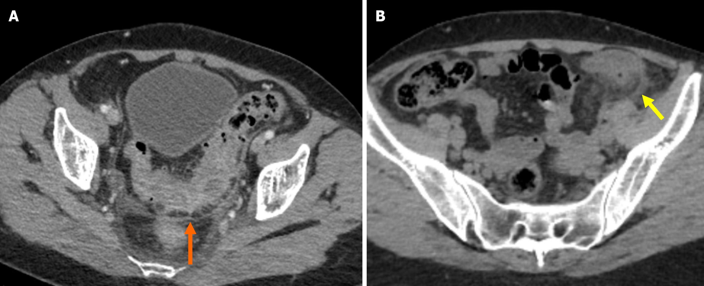
Figure 1 Acute uncomplicated diverticulitis.
A: Axial contrast-enhanced computed tomography (CT) image showed engorgement of the mesenteric vessels, a finding known as the centipede sign (orange arrow); B: Axial non-contrast CT image showed thickening and fluid collection of the left latero-conal fascia, a finding known as the comma sign (yellow arrow).
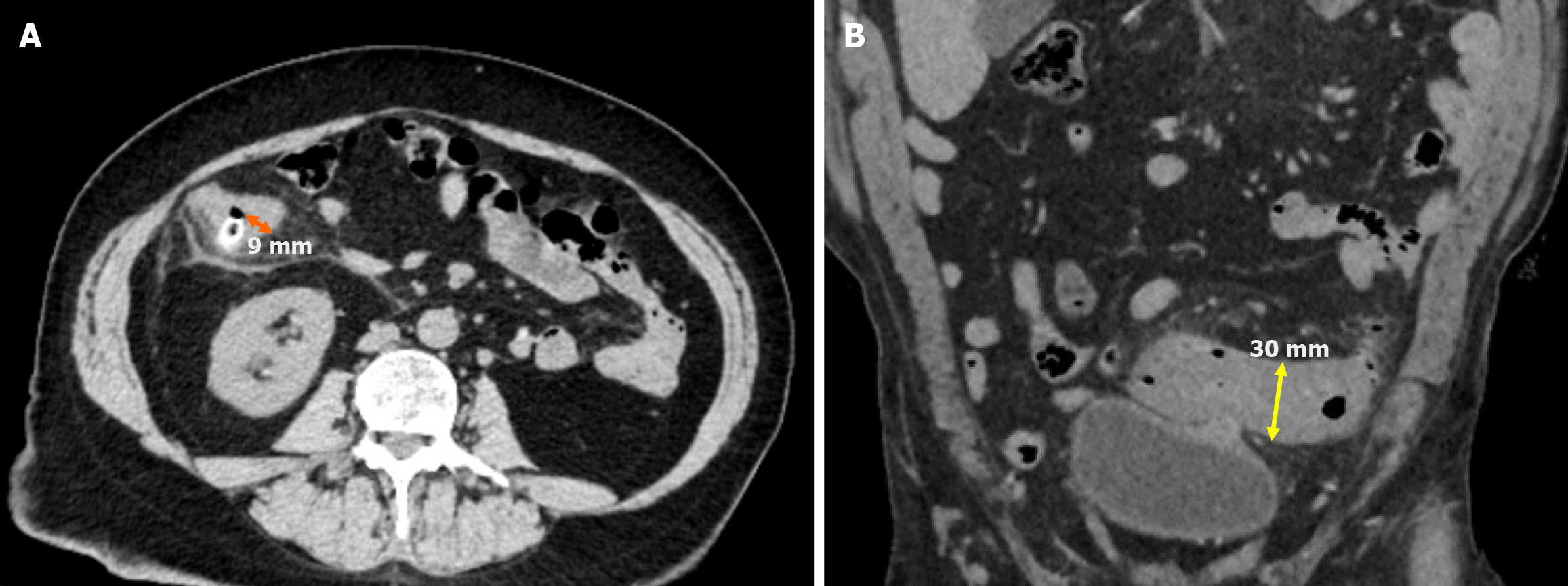
Figure 2 Measurement of colonic wall thickness.
A: Axial non-contrast computed tomography (CT) image demonstrated right colonic diverticulitis with a distinguishable lumen. The maximum colonic wall thickness was 9 mm (orange line); B: Coronal non-contrast CT image showed sigmoid colonic diverticulitis. Since the lumen was not visualized, the serosa-to-serosa distance was measured at 30 mm (yellow double-sided arrow). Half of this distance was the estimated luminal wall thickness, calculated as 15 mm.
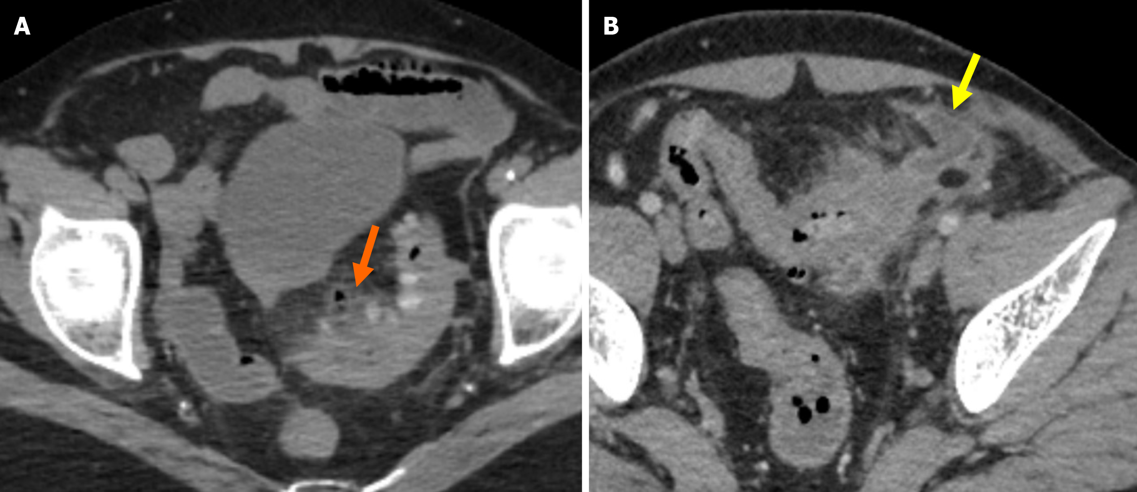
Figure 3 Sartelli stage 1A and stage 1B diverticulitis.
A: On contrast-enhanced axial computed tomography (CT) imaging, colonic wall thickening, fat stranding, and pericolic air bubbles were observed in the sigmoid colon, consistent with Sartelli stage 1A diverticulitis (orange arrow); B: Contrast-enhanced axial CT imaging revealed findings consistent with Sartelli stage 1B diverticulitis, including colonic wall thickening, pericolic fat stranding, and a fluid collection measuring approximately 2.5 cm on the anterior aspect of the sigmoid colon, indicative of an abscess (yellow arrow).
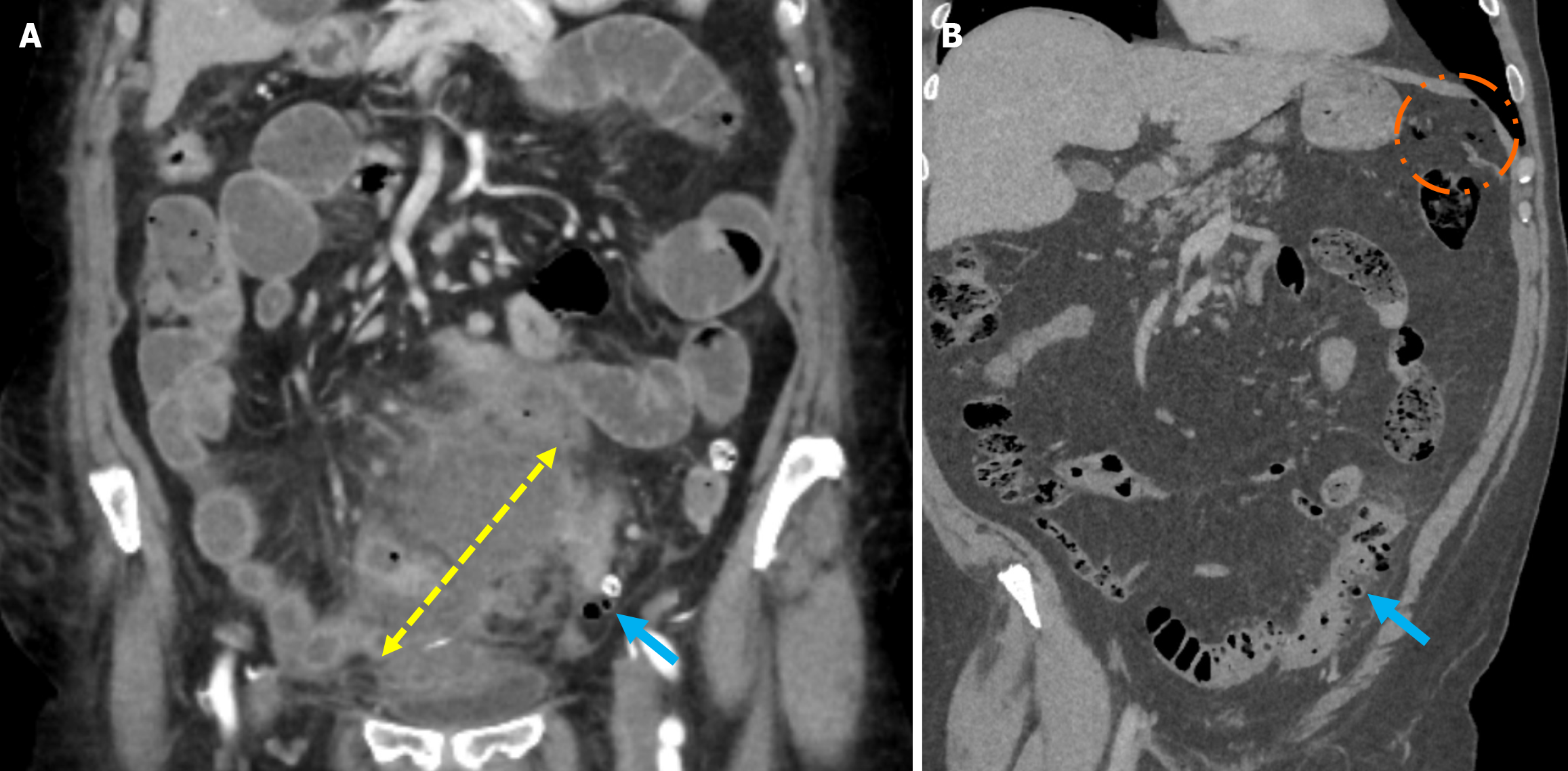
Figure 4 Sartelli stage 2A and stage 2B diverticulitis.
A: Contrast-enhanced coronal reformatted computed tomography (CT) images demonstrated diverticulitis in the sigmoid colon (blue arrow) and an adjacent abscess formation measuring approximately 7 cm long (yellow double-sided arrow), consistent with Sartelli stage 2A diverticulitis; B: Contrast-enhanced coronal CT images revealed long-segment diffuse colonic wall thickening, pericolic fat stranding, and diverticulitis in the sigmoid colon (blue arrow) along with the presence of intraperitoneal free millimetric air (orange circle), consistent with Sartelli stage 2B diverticulitis.
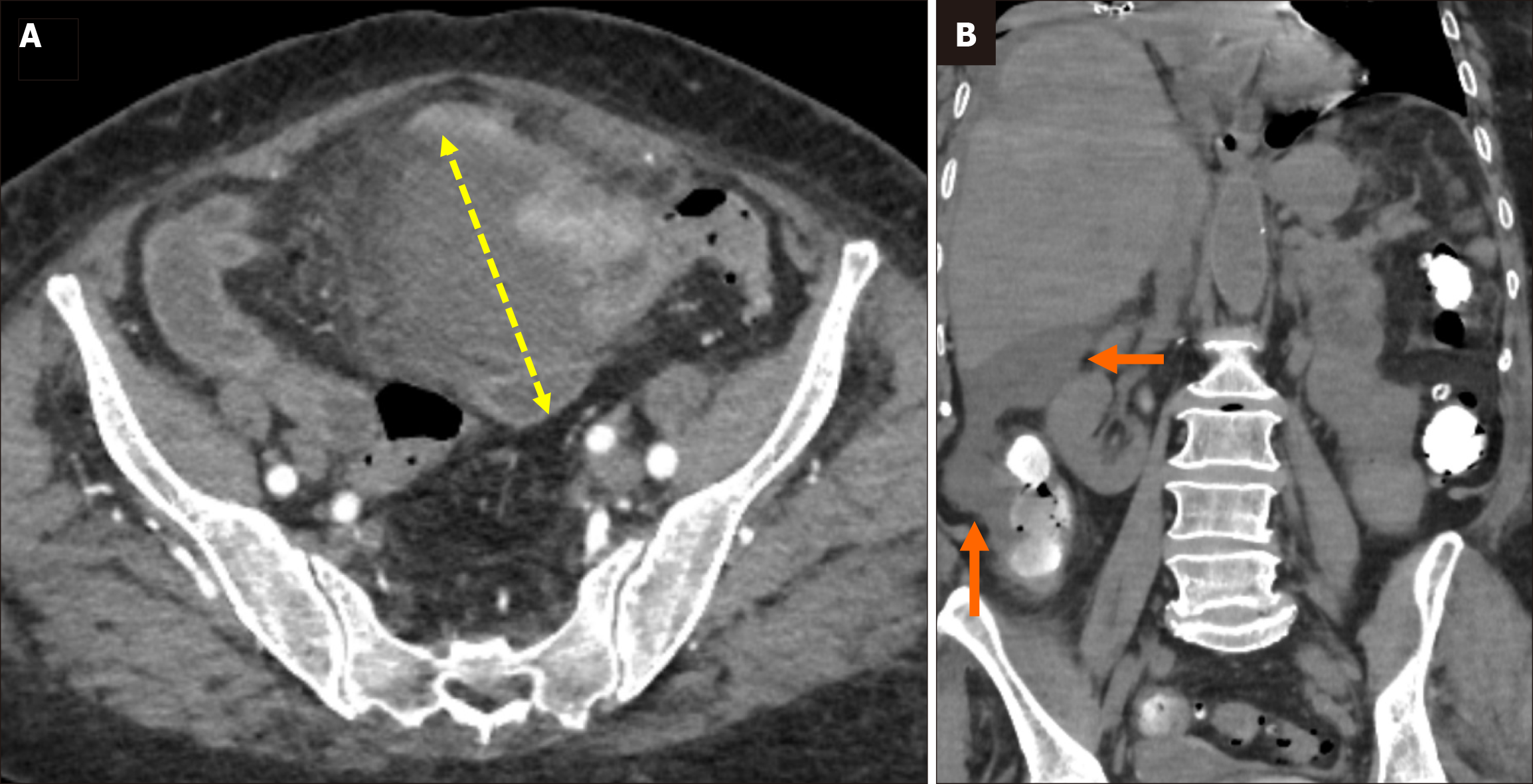
Figure 5 Sartelli stage 3 diverticulitis.
A: Axial contrast-enhanced computed tomography (CT) demonstrated findings of sigmoid colon diverticulitis with an associated abscess formation long (yellow double-sided arrow) in the midline abdomen; B: Coronal oral contrast-enhanced CT image showed free intraperitoneal fluid extending into the right paracolic and perihepatic spaces (orange arrows), with no evidence of free air in the same patient.
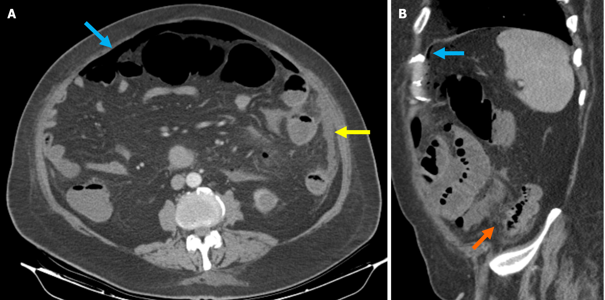
Figure 6 Sartelli stage 4 diverticulitis.
A: Axial reformatted contrast-enhanced computed tomography (CT) image including widespread intraabdominal free air (blue arrow) and free fluid (yellow arrow), more prominently around the left colon; B: Sagittal reformatted contrast-enhanced CT image also included widespread intraabdominal free air (blue arrow). Pericolic fat stranding and inflammatory changes in the pericolic fat (orange arrow) were also observed, indicative of diverticulitis. Surgical intervention confirmed perforated diverticulitis originating from the left colon.
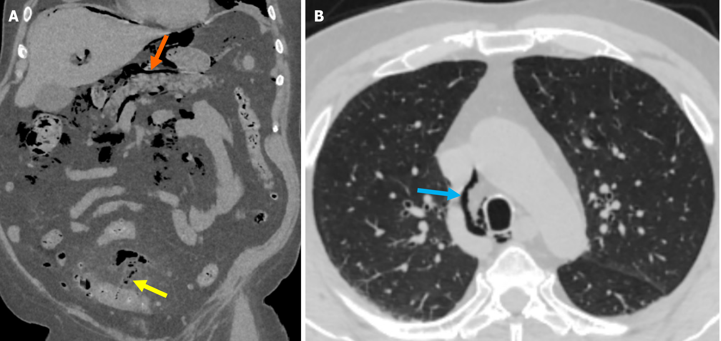
Figure 7 Free air extending to the mediastinum due to sigmoid colon diverticulitis perforation.
A: Non-contrast coronal reformatted computed tomography (CT) image at the abdominal level demonstrated the presence of air in the mesentery of the sigmoid colon (yellow arrow), consistent with perforation. The air was seen surrounding the portal structures in the hepatic hilum and extending retroperitoneally (orange arrow); B: Lung window axial CT image at the thoracic level showed that the air column extended to the mediastinum (blue arrow).
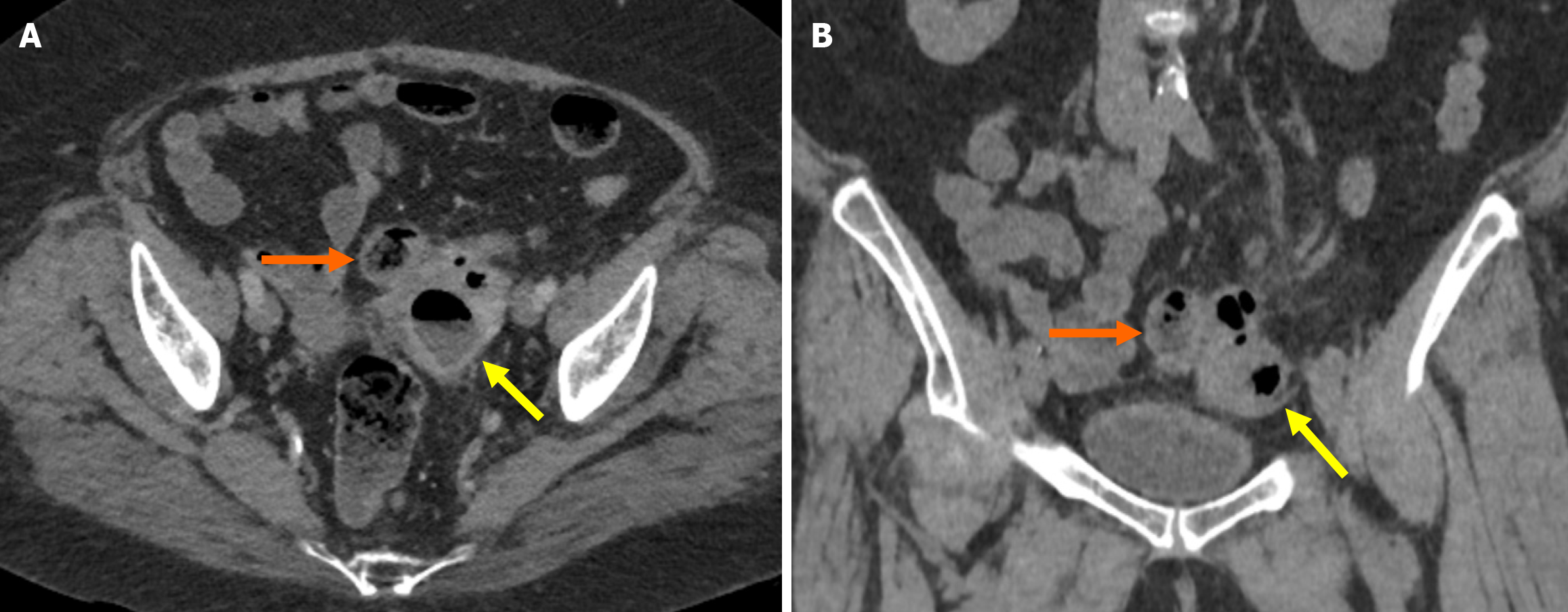
Figure 8 Tubo-ovarian abscess secondary to sigmoid colon diverticulitis.
A: Contrast-enhanced axial; B: Non-contrast coronal computed tomography images. Demonstrated a left tubo-ovarian abscess (yellow arrows) in a postmenopausal female patient with a history of diverticular disease. The abscess, which was later determined secondary to diverticulitis, was observed with a loss of fat planes between the sigmoid colon (orange arrows) and the abscess area.
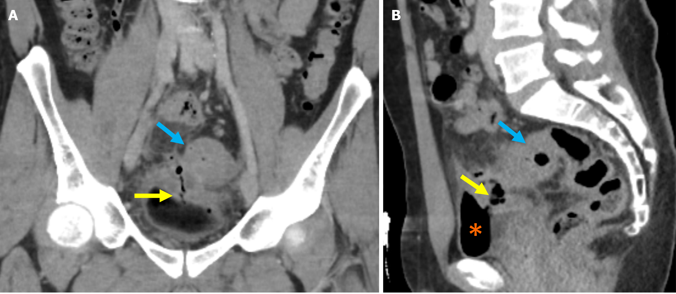
Figure 9 Colo-vesical fistula secondary to sigmoid colon diverticulitis.
A young male patient with fecaluria and pneumaturia. A: Coronal contrast-enhanced computed tomography image demonstrated findings consistent with sigmoid diverticulitis. Loss of fat planes between the sigmoid colon (blue arrows), an air-filled tract extending from the sigmoid colon to the bladder lumen, indicative of a colo-vesical fistula (yellow arrows), and the adjacent bladder segment with bladder wall thickening in the affected area was visualized; B: The presence of intraluminal air within the bladder was noted (orange asterisks).
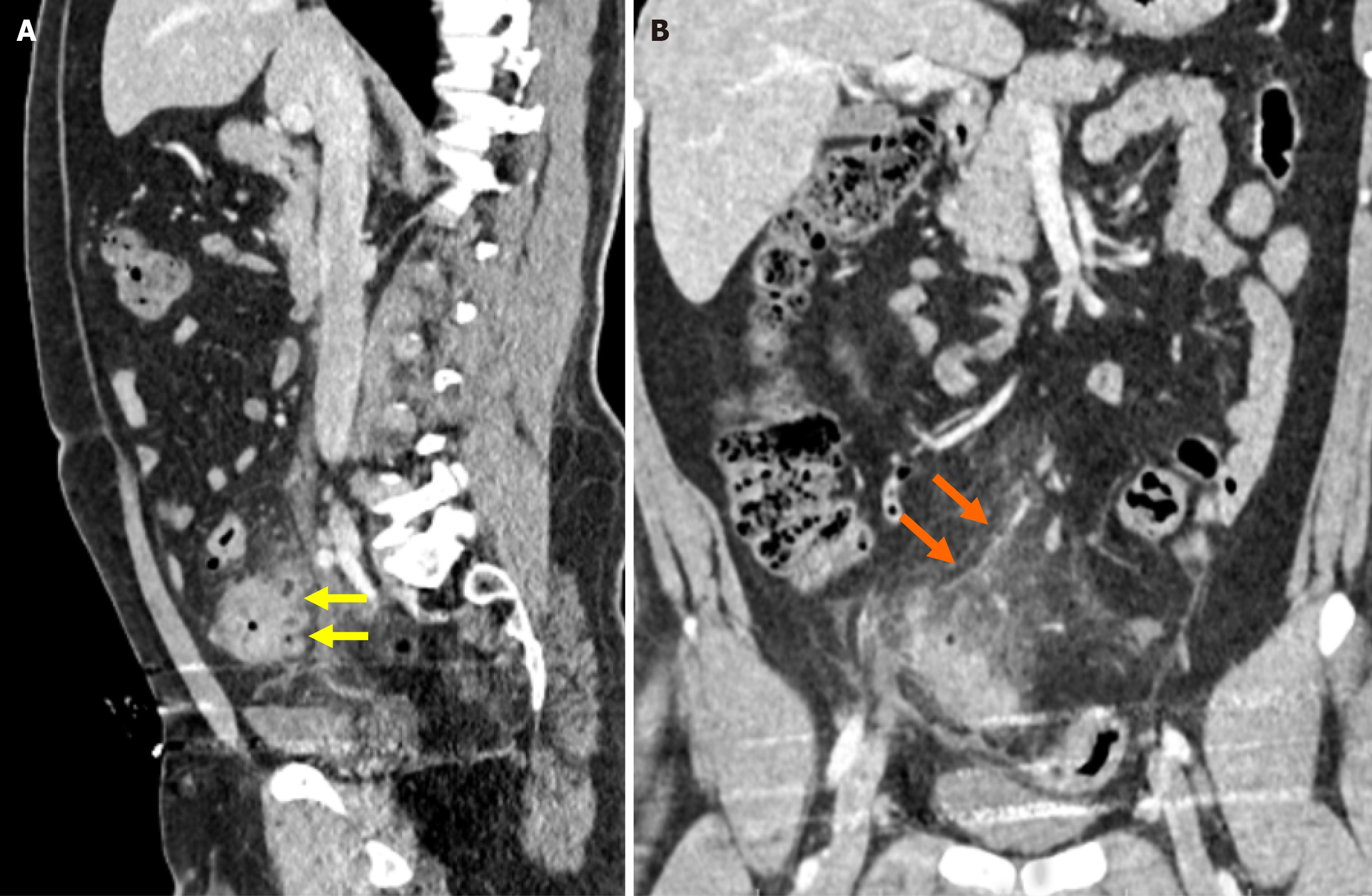
Figure 10 Pylephlebitis secondary to sigmoid colon diverticulitis.
A: Contrast-enhanced sagittal reformatted computed tomography (CT) image demonstrated segmental wall thickening of the sigmoid colon, fat stranding in the surrounding mesenteric fat, and the presence of multiple diverticula, consistent with diverticulitis (yellow arrow); B: Contrast-enhanced coronal reformatted CT image showed a dilated venous structure with a lack of contrast enhancement in continuity with the inferior mesenteric vein, suggestive of thrombophlebitis (orange arrow).
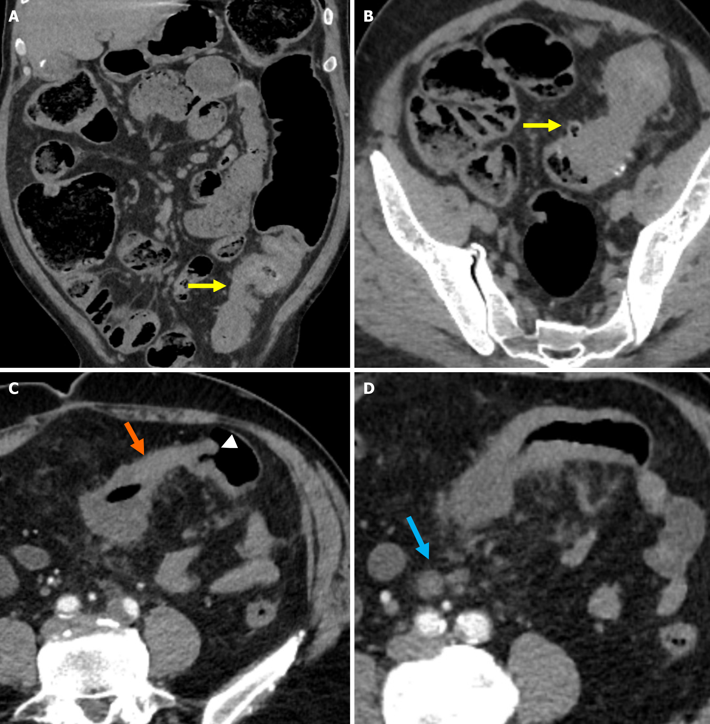
Figure 11 Distinguishing features of chronic sigmoid diverticulitis and colorectal cancer.
A and B: Non-contrast-enhanced coronal reformatted computed tomography (CT) image and non-contrast-enhanced axial CT image showed long-segment, diffuse wall thickening in the sigmoid colon, luminal narrowing due to fibrosis, and diverticula (yellow arrows). Recurrent obstruction with proximal bowel loop dilatation was evident. The absence of pathological lymphadenopathy in the mesocolon served as a key diagnostic clue; C: Marked wall thickening of the affected bowel segment (orange arrow) with the shoulder phenomenon (white arrowhead) without visible diverticula; D: Rounded, metastatic lymph nodes (blue arrow) in the mesentery of the affected sigmoid segment accompanied by fat stranding consistent with pericolic fat infiltration.
- Citation: Simsar M, Yuruk YY, Sahin O, Sahin H. Radiological insights into diverticulitis: Clinical manifestations, complications, and differential diagnosis. World J Radiol 2025; 17(8): 107463
- URL: https://www.wjgnet.com/1949-8470/full/v17/i8/107463.htm
- DOI: https://dx.doi.org/10.4329/wjr.v17.i8.107463


