Copyright
©The Author(s) 2025.
World J Radiol. Aug 28, 2025; 17(8): 107463
Published online Aug 28, 2025. doi: 10.4329/wjr.v17.i8.107463
Published online Aug 28, 2025. doi: 10.4329/wjr.v17.i8.107463
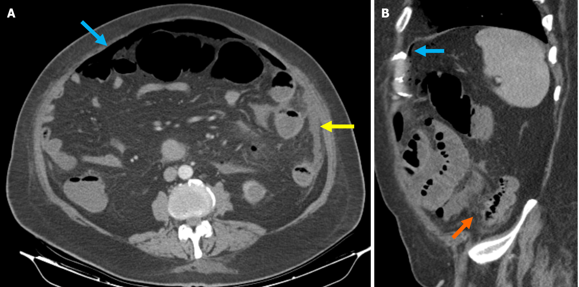
Figure 6 Sartelli stage 4 diverticulitis.
A: Axial reformatted contrast-enhanced computed tomography (CT) image including widespread intraabdominal free air (blue arrow) and free fluid (yellow arrow), more prominently around the left colon; B: Sagittal reformatted contrast-enhanced CT image also included widespread intraabdominal free air (blue arrow). Pericolic fat stranding and inflammatory changes in the pericolic fat (orange arrow) were also observed, indicative of diverticulitis. Surgical intervention confirmed perforated diverticulitis originating from the left colon.
- Citation: Simsar M, Yuruk YY, Sahin O, Sahin H. Radiological insights into diverticulitis: Clinical manifestations, complications, and differential diagnosis. World J Radiol 2025; 17(8): 107463
- URL: https://www.wjgnet.com/1949-8470/full/v17/i8/107463.htm
- DOI: https://dx.doi.org/10.4329/wjr.v17.i8.107463


