
















©The Author(s) 2025.
World J Radiol. Aug 28, 2025; 17(8): 107463
Published online Aug 28, 2025. doi: 10.4329/wjr.v17.i8.107463
Published online Aug 28, 2025. doi: 10.4329/wjr.v17.i8.107463
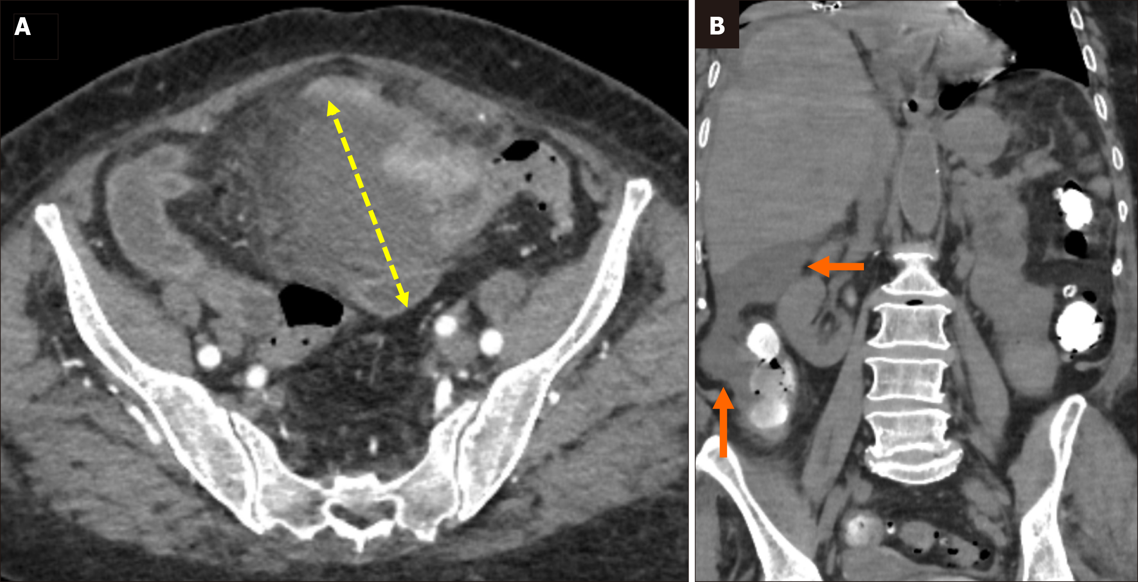
Figure 5 Sartelli stage 3 diverticulitis.
A: Axial contrast-enhanced computed tomography (CT) demonstrated findings of sigmoid colon diverticulitis with an associated abscess formation long (yellow double-sided arrow) in the midline abdomen; B: Coronal oral contrast-enhanced CT image showed free intraperitoneal fluid extending into the right paracolic and perihepatic spaces (orange arrows), with no evidence of free air in the same patient.
- Citation: Simsar M, Yuruk YY, Sahin O, Sahin H. Radiological insights into diverticulitis: Clinical manifestations, complications, and differential diagnosis. World J Radiol 2025; 17(8): 107463
- URL: https://www.wjgnet.com/1949-8470/full/v17/i8/107463.htm
- DOI: https://dx.doi.org/10.4329/wjr.v17.i8.107463