Copyright
©The Author(s) 2025.
World J Radiol. Aug 28, 2025; 17(8): 107463
Published online Aug 28, 2025. doi: 10.4329/wjr.v17.i8.107463
Published online Aug 28, 2025. doi: 10.4329/wjr.v17.i8.107463
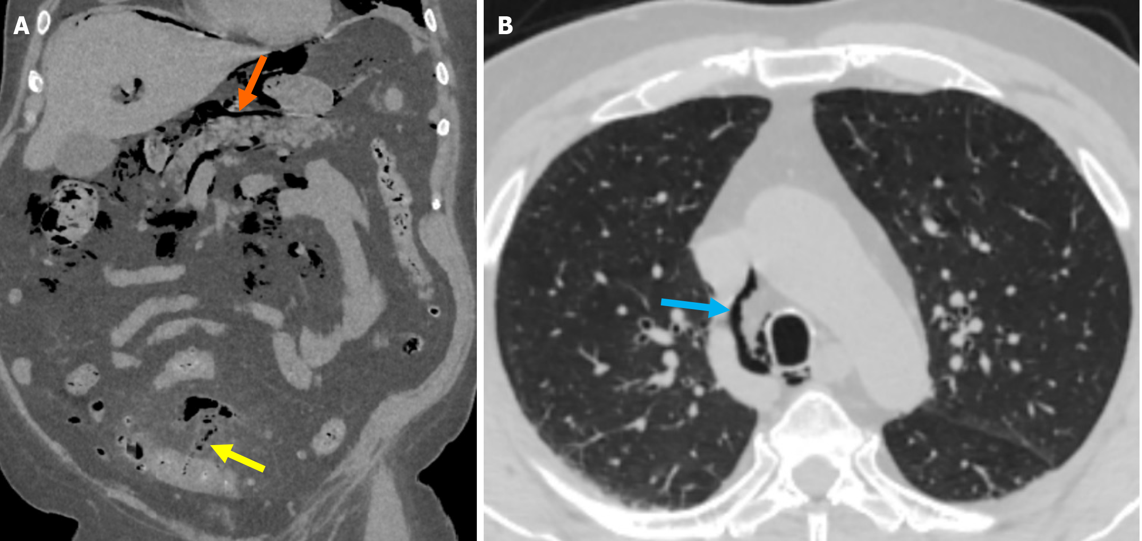
Figure 7 Free air extending to the mediastinum due to sigmoid colon diverticulitis perforation.
A: Non-contrast coronal reformatted computed tomography (CT) image at the abdominal level demonstrated the presence of air in the mesentery of the sigmoid colon (yellow arrow), consistent with perforation. The air was seen surrounding the portal structures in the hepatic hilum and extending retroperitoneally (orange arrow); B: Lung window axial CT image at the thoracic level showed that the air column extended to the mediastinum (blue arrow).
- Citation: Simsar M, Yuruk YY, Sahin O, Sahin H. Radiological insights into diverticulitis: Clinical manifestations, complications, and differential diagnosis. World J Radiol 2025; 17(8): 107463
- URL: https://www.wjgnet.com/1949-8470/full/v17/i8/107463.htm
- DOI: https://dx.doi.org/10.4329/wjr.v17.i8.107463


