Copyright
©The Author(s) 2025.
World J Radiol. Aug 28, 2025; 17(8): 107463
Published online Aug 28, 2025. doi: 10.4329/wjr.v17.i8.107463
Published online Aug 28, 2025. doi: 10.4329/wjr.v17.i8.107463
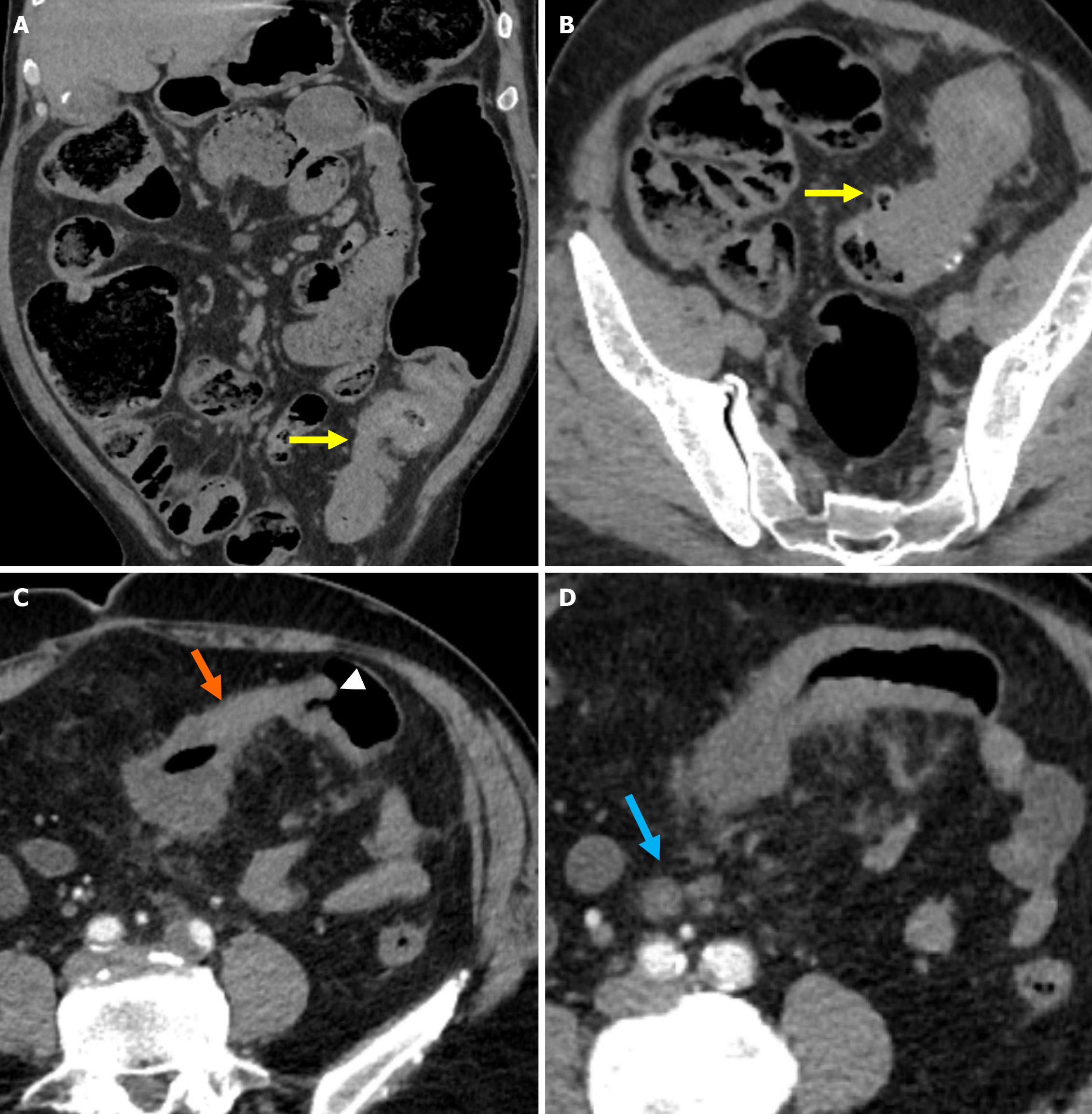
Figure 11 Distinguishing features of chronic sigmoid diverticulitis and colorectal cancer.
A and B: Non-contrast-enhanced coronal reformatted computed tomography (CT) image and non-contrast-enhanced axial CT image showed long-segment, diffuse wall thickening in the sigmoid colon, luminal narrowing due to fibrosis, and diverticula (yellow arrows). Recurrent obstruction with proximal bowel loop dilatation was evident. The absence of pathological lymphadenopathy in the mesocolon served as a key diagnostic clue; C: Marked wall thickening of the affected bowel segment (orange arrow) with the shoulder phenomenon (white arrowhead) without visible diverticula; D: Rounded, metastatic lymph nodes (blue arrow) in the mesentery of the affected sigmoid segment accompanied by fat stranding consistent with pericolic fat infiltration.
- Citation: Simsar M, Yuruk YY, Sahin O, Sahin H. Radiological insights into diverticulitis: Clinical manifestations, complications, and differential diagnosis. World J Radiol 2025; 17(8): 107463
- URL: https://www.wjgnet.com/1949-8470/full/v17/i8/107463.htm
- DOI: https://dx.doi.org/10.4329/wjr.v17.i8.107463


