Copyright
©The Author(s) 2025.
World J Radiol. Aug 28, 2025; 17(8): 107463
Published online Aug 28, 2025. doi: 10.4329/wjr.v17.i8.107463
Published online Aug 28, 2025. doi: 10.4329/wjr.v17.i8.107463
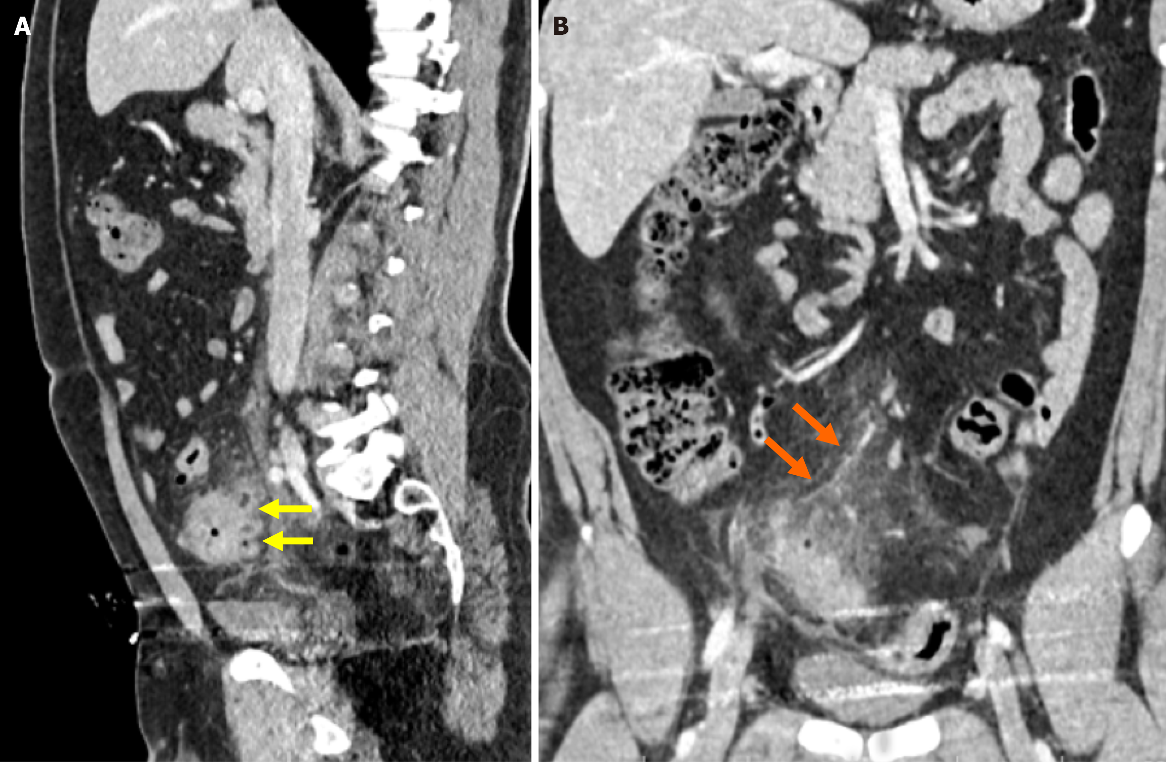
Figure 10 Pylephlebitis secondary to sigmoid colon diverticulitis.
A: Contrast-enhanced sagittal reformatted computed tomography (CT) image demonstrated segmental wall thickening of the sigmoid colon, fat stranding in the surrounding mesenteric fat, and the presence of multiple diverticula, consistent with diverticulitis (yellow arrow); B: Contrast-enhanced coronal reformatted CT image showed a dilated venous structure with a lack of contrast enhancement in continuity with the inferior mesenteric vein, suggestive of thrombophlebitis (orange arrow).
- Citation: Simsar M, Yuruk YY, Sahin O, Sahin H. Radiological insights into diverticulitis: Clinical manifestations, complications, and differential diagnosis. World J Radiol 2025; 17(8): 107463
- URL: https://www.wjgnet.com/1949-8470/full/v17/i8/107463.htm
- DOI: https://dx.doi.org/10.4329/wjr.v17.i8.107463


