Copyright
©The Author(s) 2025.
World J Radiol. Aug 28, 2025; 17(8): 107463
Published online Aug 28, 2025. doi: 10.4329/wjr.v17.i8.107463
Published online Aug 28, 2025. doi: 10.4329/wjr.v17.i8.107463
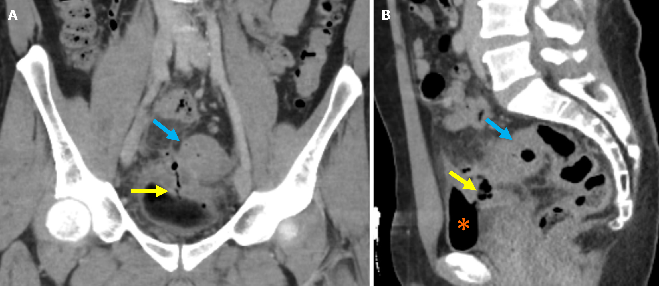
Figure 9 Colo-vesical fistula secondary to sigmoid colon diverticulitis.
A young male patient with fecaluria and pneumaturia. A: Coronal contrast-enhanced computed tomography image demonstrated findings consistent with sigmoid diverticulitis. Loss of fat planes between the sigmoid colon (blue arrows), an air-filled tract extending from the sigmoid colon to the bladder lumen, indicative of a colo-vesical fistula (yellow arrows), and the adjacent bladder segment with bladder wall thickening in the affected area was visualized; B: The presence of intraluminal air within the bladder was noted (orange asterisks).
- Citation: Simsar M, Yuruk YY, Sahin O, Sahin H. Radiological insights into diverticulitis: Clinical manifestations, complications, and differential diagnosis. World J Radiol 2025; 17(8): 107463
- URL: https://www.wjgnet.com/1949-8470/full/v17/i8/107463.htm
- DOI: https://dx.doi.org/10.4329/wjr.v17.i8.107463


