Copyright
©The Author(s) 2025.
World J Radiol. Aug 28, 2025; 17(8): 107463
Published online Aug 28, 2025. doi: 10.4329/wjr.v17.i8.107463
Published online Aug 28, 2025. doi: 10.4329/wjr.v17.i8.107463
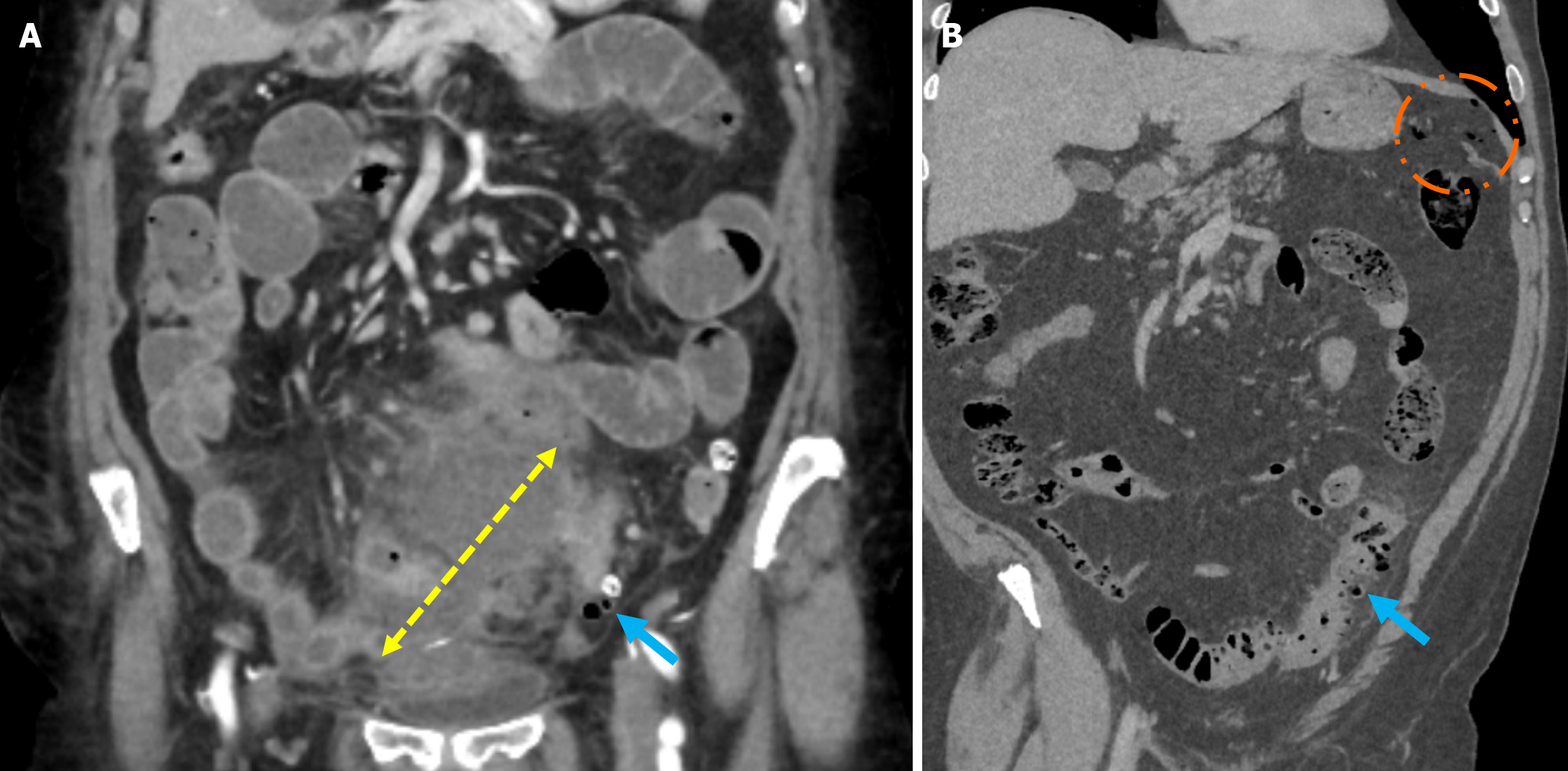
Figure 4 Sartelli stage 2A and stage 2B diverticulitis.
A: Contrast-enhanced coronal reformatted computed tomography (CT) images demonstrated diverticulitis in the sigmoid colon (blue arrow) and an adjacent abscess formation measuring approximately 7 cm long (yellow double-sided arrow), consistent with Sartelli stage 2A diverticulitis; B: Contrast-enhanced coronal CT images revealed long-segment diffuse colonic wall thickening, pericolic fat stranding, and diverticulitis in the sigmoid colon (blue arrow) along with the presence of intraperitoneal free millimetric air (orange circle), consistent with Sartelli stage 2B diverticulitis.
- Citation: Simsar M, Yuruk YY, Sahin O, Sahin H. Radiological insights into diverticulitis: Clinical manifestations, complications, and differential diagnosis. World J Radiol 2025; 17(8): 107463
- URL: https://www.wjgnet.com/1949-8470/full/v17/i8/107463.htm
- DOI: https://dx.doi.org/10.4329/wjr.v17.i8.107463


