Copyright
©The Author(s) 2025.
World J Radiol. Aug 28, 2025; 17(8): 107463
Published online Aug 28, 2025. doi: 10.4329/wjr.v17.i8.107463
Published online Aug 28, 2025. doi: 10.4329/wjr.v17.i8.107463
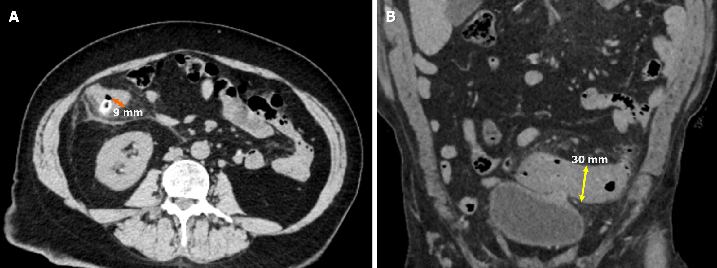
Figure 2 Measurement of colonic wall thickness.
A: Axial non-contrast computed tomography (CT) image demonstrated right colonic diverticulitis with a distinguishable lumen. The maximum colonic wall thickness was 9 mm (orange line); B: Coronal non-contrast CT image showed sigmoid colonic diverticulitis. Since the lumen was not visualized, the serosa-to-serosa distance was measured at 30 mm (yellow double-sided arrow). Half of this distance was the estimated luminal wall thickness, calculated as 15 mm.
- Citation: Simsar M, Yuruk YY, Sahin O, Sahin H. Radiological insights into diverticulitis: Clinical manifestations, complications, and differential diagnosis. World J Radiol 2025; 17(8): 107463
- URL: https://www.wjgnet.com/1949-8470/full/v17/i8/107463.htm
- DOI: https://dx.doi.org/10.4329/wjr.v17.i8.107463


