Copyright
©The Author(s) 2022.
World J Clin Cases. Jul 6, 2022; 10(19): 6750-6758
Published online Jul 6, 2022. doi: 10.12998/wjcc.v10.i19.6750
Published online Jul 6, 2022. doi: 10.12998/wjcc.v10.i19.6750
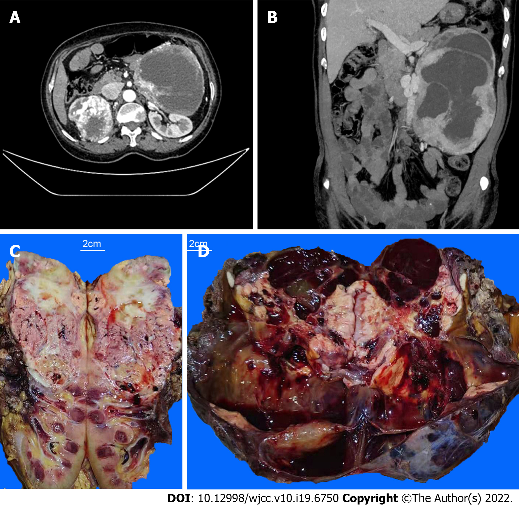
Figure 1 Enhance computed tomography scan of the upper and lower abdomen and the cut surfaces of tumor masses.
A: A tumor mass in the patient’s right kidney; B: A tumor mass in her left retroperitoneum; C: The upper portion of the patient’s right kidney was replaced by a tumor mass. The cutting section of the tumor was tan to whitish or yellowish solid, with hemorrhage and necrosis; D: The cutting section of the left retroperitoneal mass was dark red with cystic changes, hemorrhage, and necrosis. Distinct whitish to yellowish confluenting tumor nodules were observed within the major mass.
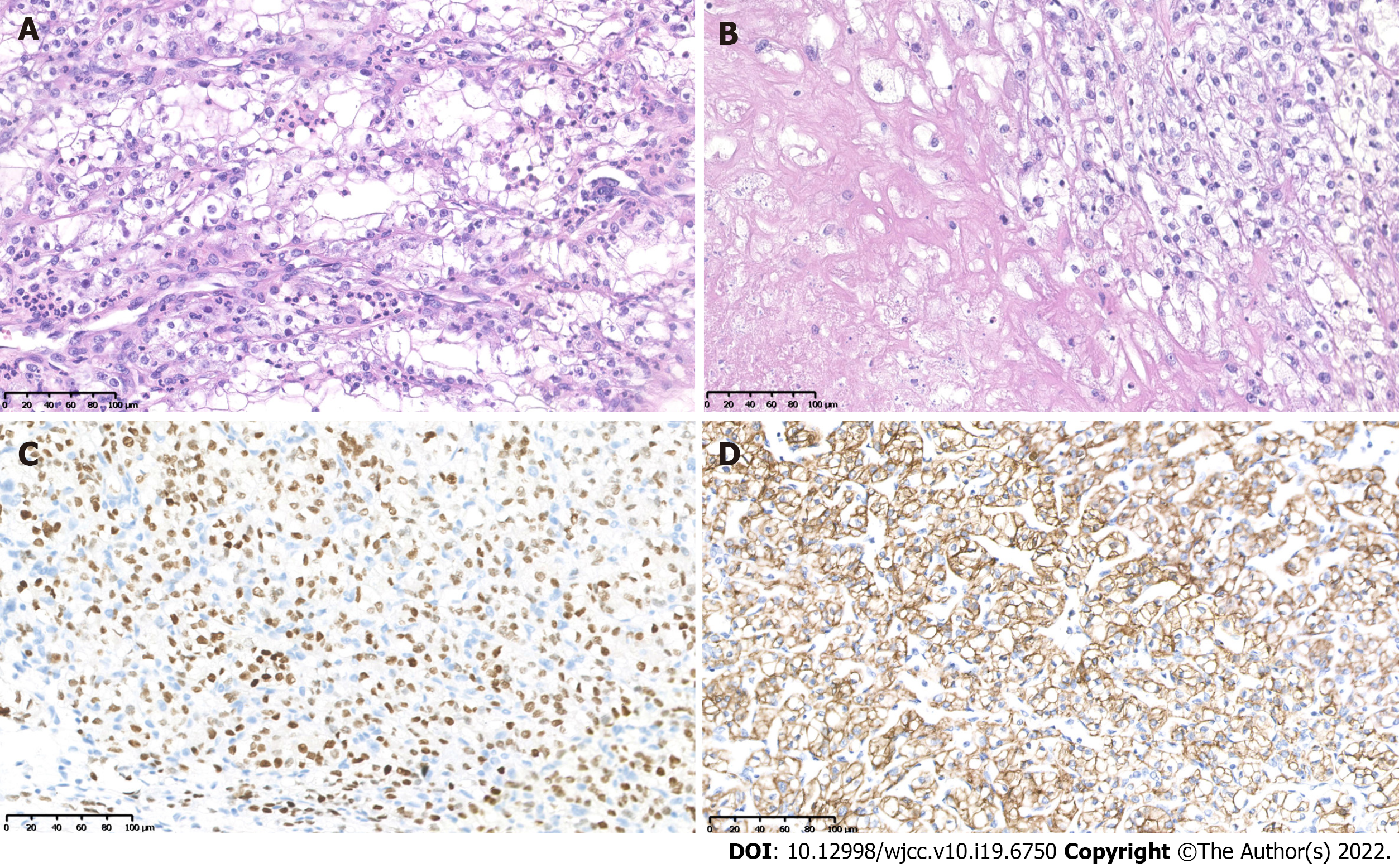
Figure 2 Histopathological and immunohistochemiscal figures of the right kidney.
A: Typical clear cell renal cell carcinoma morphology of the right kidney tumor; B: Necrosis in the right kidney tumor; C: The tumor showed positive for PAX8; D: The tumor showed positive for CAIX.
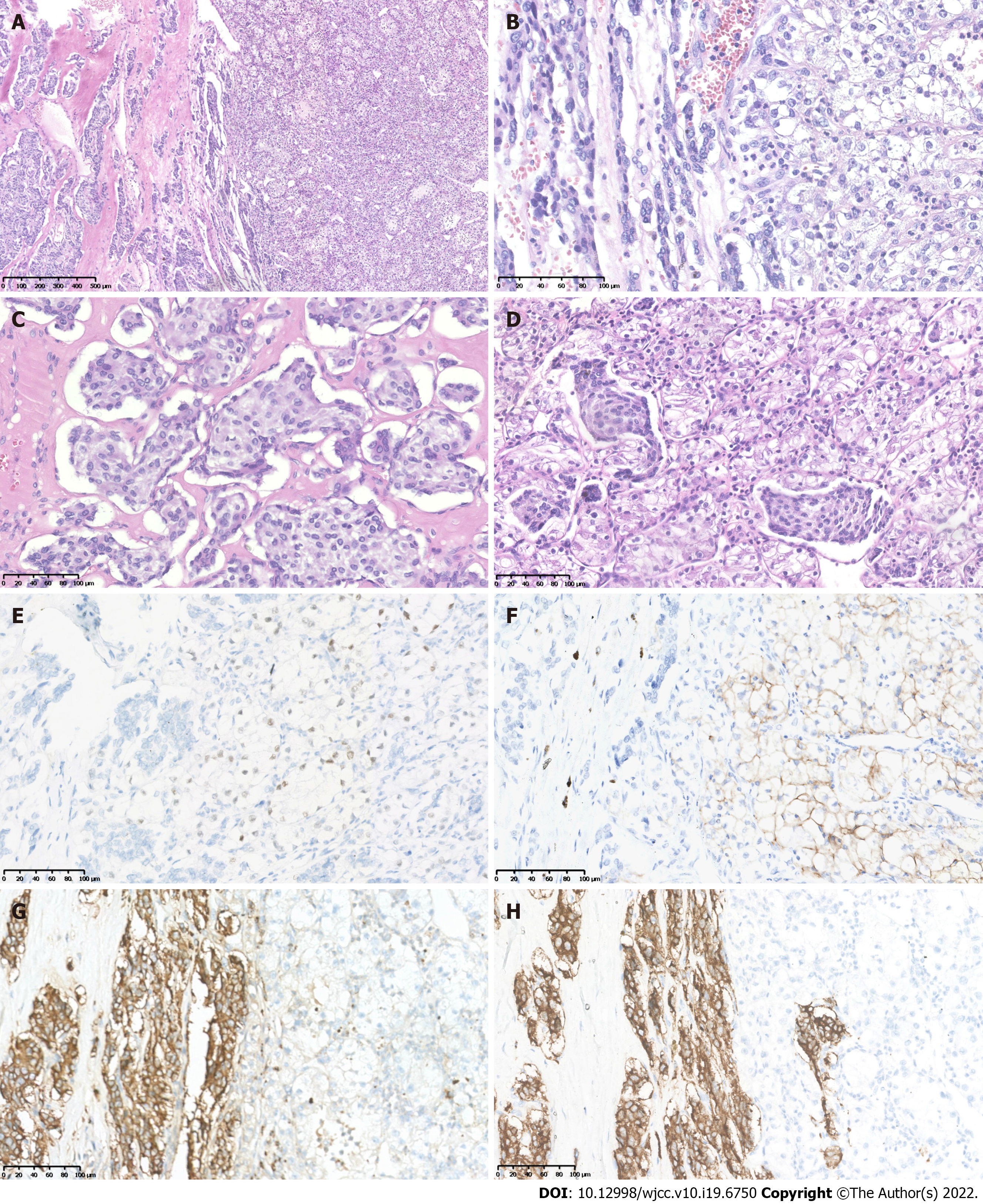
Figure 3 Histopathological and immunohistochemiscal figures showed the left retroperitoneal tumor mass contained two components.
A: One component was typical pheochromocytoma (PCC) with tumor cells arranged in organoid nests and cords; B: The other component exhibited clear cell renal cell carcinoma (CCRCC) morphology; C: The PCC nests at high magnification; D: The PCC nests mixed with CCRCC at their intersection; E: Immunohistochemistry (IHC) showed PAX8 was positive in the CCRCC tumor cells; F: IHC showed CAIX was positive in the CCRCC tumor cells; G: PCC tumor cells showed positive for CgA; H: PCC tumor cells showed positive for Syn.
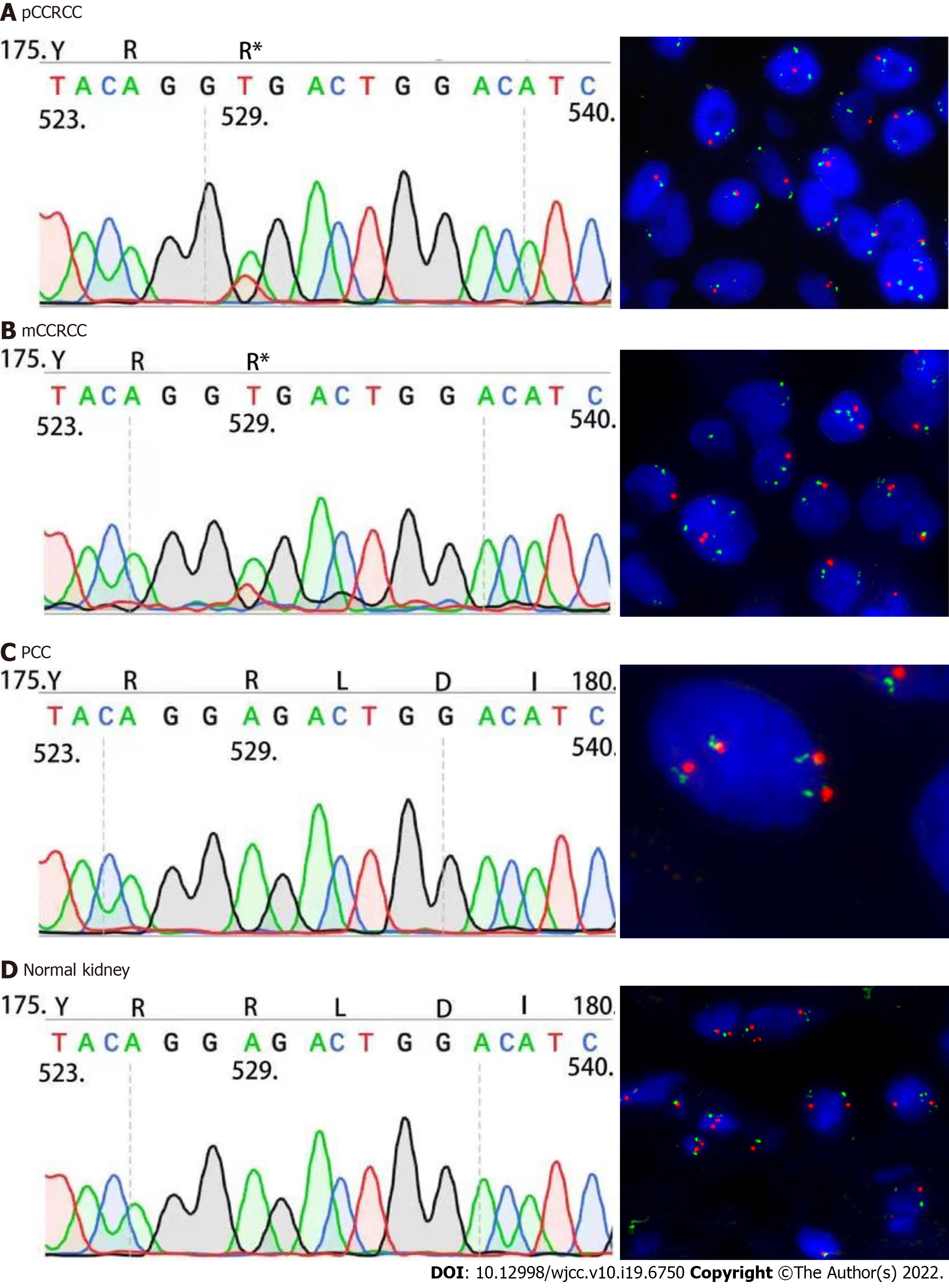
Figure 4 Results of Sanger sequencing and fluorescence in situ hybridization.
A: Sanger sequencing showed identical VHL c.529A>T mutation and fluorescence in situ hybridization analysis showed the loss of 3p in the right kidney clear cell renal cell carcinoma (CCRCC); B: The VHL c.529A>T mutation and loss of 3p in metastatic CCRCC component within the left pheochromocytoma (PCC); C: The left retroperitoneal PCC did not carry VHL c.529A>T mutation and loss of 3p; D: The right normal kidney tissue did not carry VHL c.529A>T mutation and loss of 3p. CCRCC: Clear cell renal cell carcinoma; PCC: Pheochromocytoma.
- Citation: Wen HY, Hou J, Zeng H, Zhou Q, Chen N. Tumor-to-tumor metastasis of clear cell renal cell carcinoma to contralateral synchronous pheochromocytoma: A case report. World J Clin Cases 2022; 10(19): 6750-6758
- URL: https://www.wjgnet.com/2307-8960/full/v10/i19/6750.htm
- DOI: https://dx.doi.org/10.12998/wjcc.v10.i19.6750


