Copyright
©The Author(s) 2022.
World J Clin Cases. Jul 6, 2022; 10(19): 6750-6758
Published online Jul 6, 2022. doi: 10.12998/wjcc.v10.i19.6750
Published online Jul 6, 2022. doi: 10.12998/wjcc.v10.i19.6750
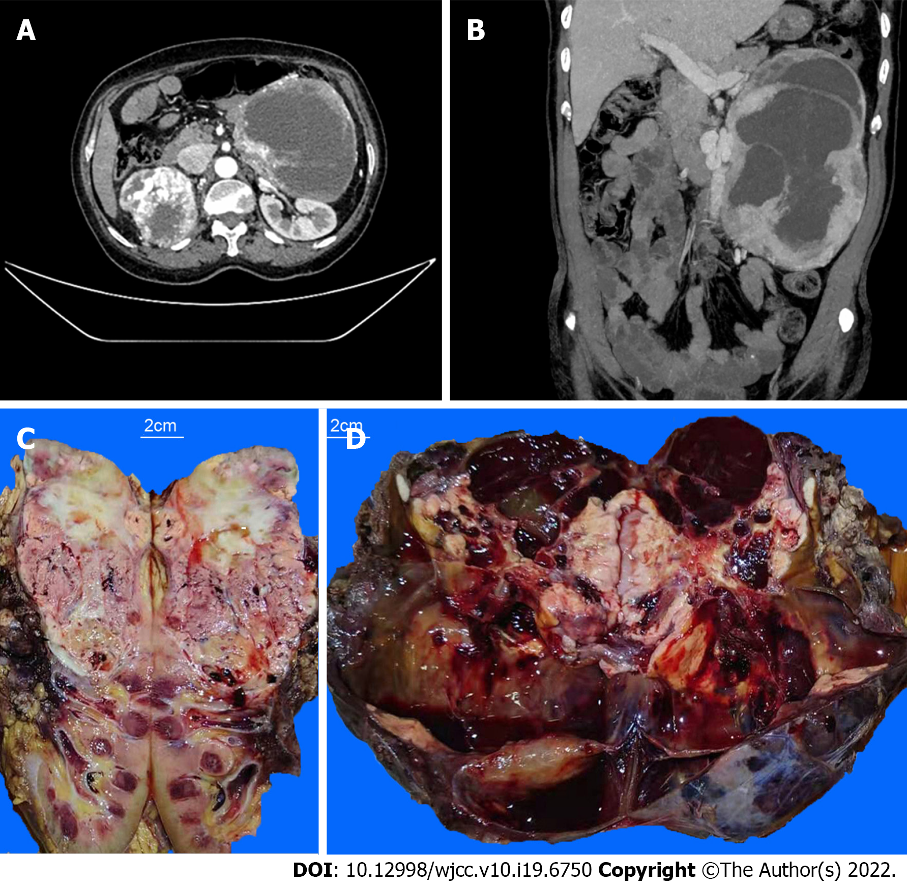
Figure 1 Enhance computed tomography scan of the upper and lower abdomen and the cut surfaces of tumor masses.
A: A tumor mass in the patient’s right kidney; B: A tumor mass in her left retroperitoneum; C: The upper portion of the patient’s right kidney was replaced by a tumor mass. The cutting section of the tumor was tan to whitish or yellowish solid, with hemorrhage and necrosis; D: The cutting section of the left retroperitoneal mass was dark red with cystic changes, hemorrhage, and necrosis. Distinct whitish to yellowish confluenting tumor nodules were observed within the major mass.
- Citation: Wen HY, Hou J, Zeng H, Zhou Q, Chen N. Tumor-to-tumor metastasis of clear cell renal cell carcinoma to contralateral synchronous pheochromocytoma: A case report. World J Clin Cases 2022; 10(19): 6750-6758
- URL: https://www.wjgnet.com/2307-8960/full/v10/i19/6750.htm
- DOI: https://dx.doi.org/10.12998/wjcc.v10.i19.6750


