
















Copyright: ©Author(s) 2026.
World J Gastrointest Oncol. Nov 15, 2026; 18(11): 124061
Published online Nov 15, 2026. doi: 10.4251/wjgo.124061
Published online Nov 15, 2026. doi: 10.4251/wjgo.124061
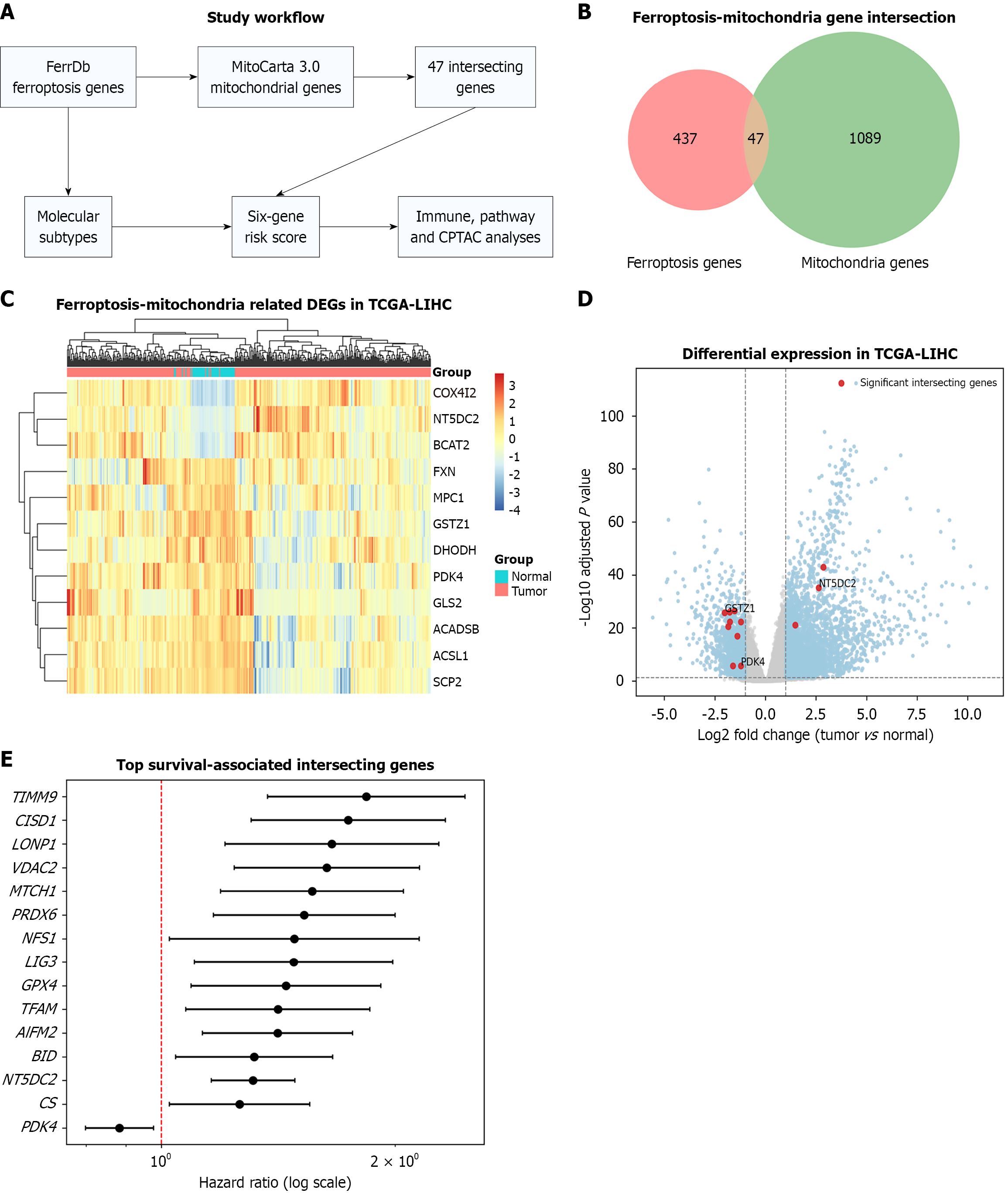
Figure 1 Identification of ferroptosis-mitochondria-related genes in hepatocellular carcinoma.
A: Study workflow; B: Venn diagram showing the overlap between ferroptosis-related and mitochondrial genes; C: Expression heatmap of the 12 significantly differentially expressed ferroptosis-mitochondria-related genes in tumor and normal liver tissues from The Cancer Genome Atlas liver hepatocellular carcinoma cohort; D: Differential expression volcano plot; E: Forest plot of representative survival-associated overlapping genes based on univariate Cox regression. TCGA-LIHC: The Cancer Genome Atlas liver hepatocellular carcinoma.
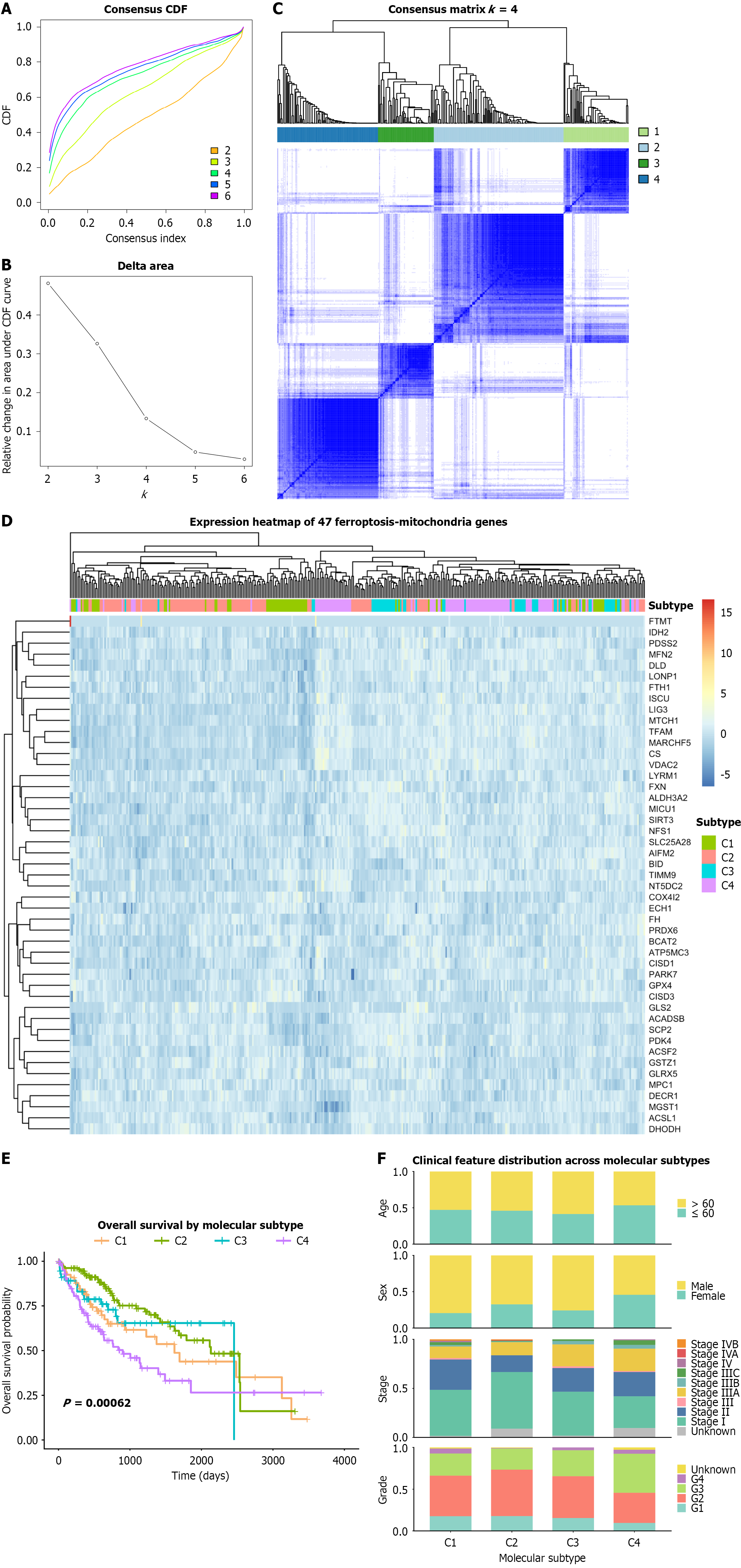
Figure 2 Identification of ferroptosis-mitochondria-related molecular subtypes in The Cancer Genome Atlas liver hepatocellular carci
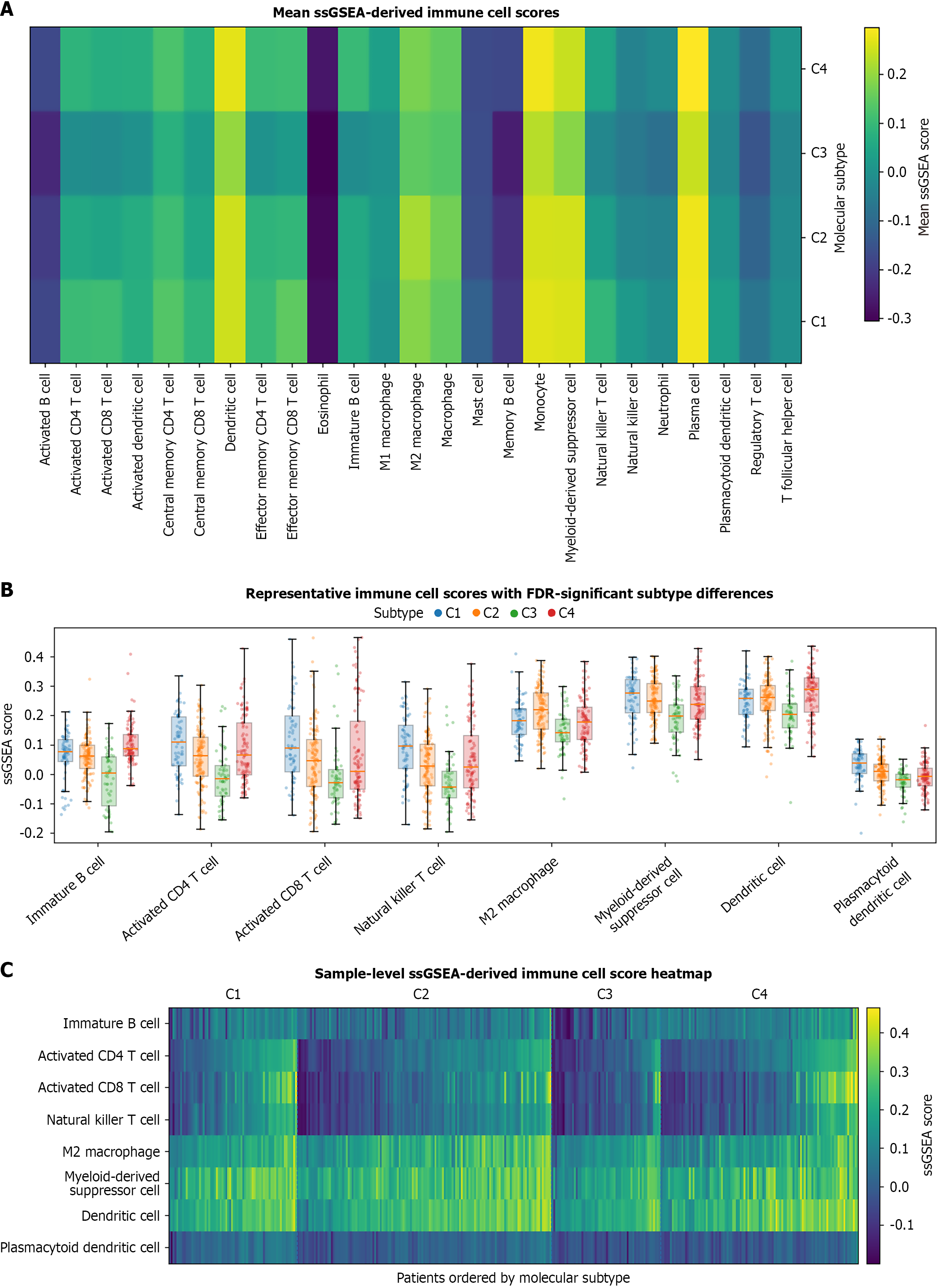
Figure 3 Single-sample gene set enrichment analysis-derived immune cell score patterns among the four molecular subtypes.
A: Mean scores of 25 immune cell populations across the four molecular subtypes; B: Boxplots with individual patient observations for representative immune cell scores showing significant subtype differences after Benjamini-Hochberg correction; C: Sample-level heatmap of representative immune cell scores ordered by molecular subtype. ssGSEA: Single-sample gene set enrichment analysis; FDR: False discovery rate.
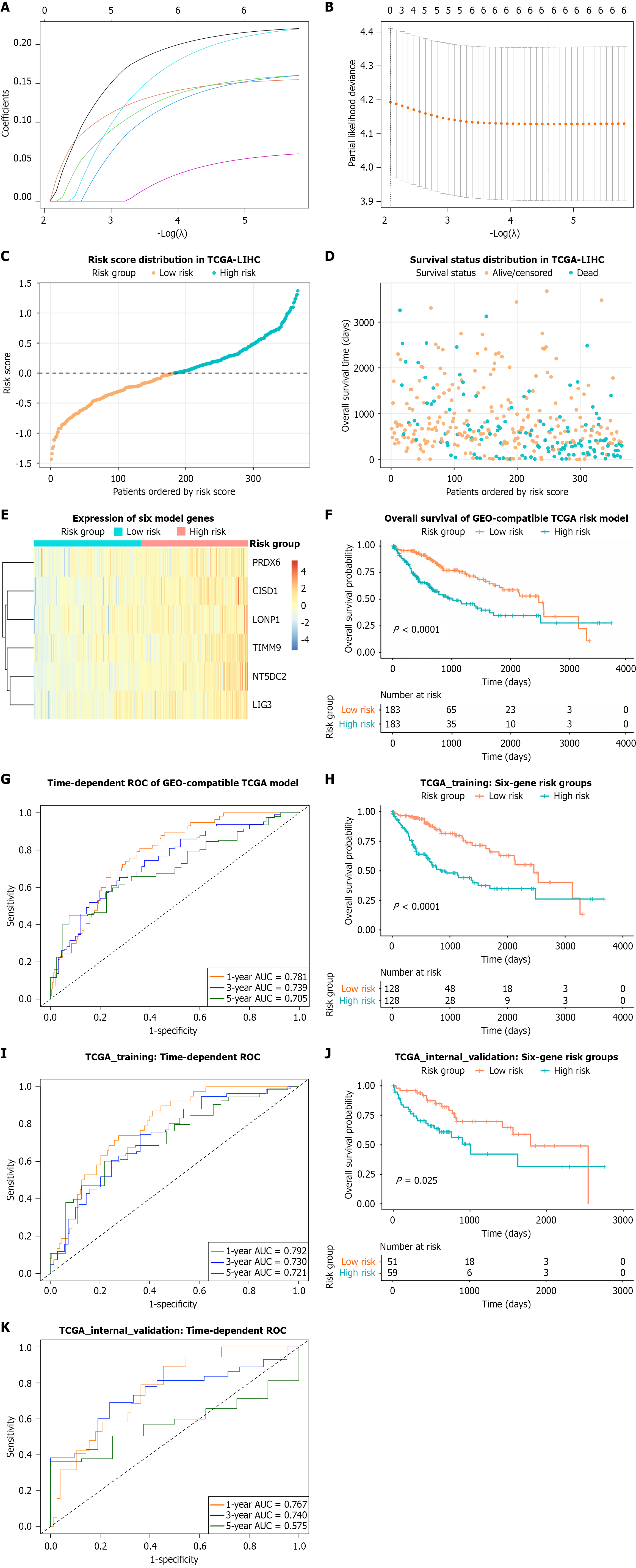
Figure 4 Construction and internal validation of the six-gene ferroptosis-mitochondria-related prognostic signature.
A: Least absolute shrinkage and selection operator coefficient profiles; B: Tenfold cross-validation curve for selection of the optimal penalty parameter; C: Risk score distribution in the full The Cancer Genome Atlas liver hepatocellular carcinoma (TCGA-LIHC) cohort; D: Survival status distribution in the full cohort; E: Heatmap of the six model genes ordered by risk score; F: Kaplan-Meier overall survival curves for the full TCGA-LIHC high- and low-risk groups; G: Time-dependent receiver operating characteristic curves for 1-, 3-, and 5-year overall survival in the full TCGA-LIHC cohort; H: Kaplan-Meier curves for the training cohort (n = 256; log-rank P = 3.66 × 10⁻⁶); I: Time-dependent receiver operating characteristic curves in the training cohort, with 1-, 3-, and 5-year areas under the curve of 0.792, 0.730, and 0.721, respectively; J: Kaplan-Meier curves for the internal validation cohort (n = 110; log-rank P = 0.0245); K: Time-dependent receiver operating characteristic curves in the internal validation cohort, with 1-, 3-, and 5-year areas under the curve of 0.767, 0.740, and 0.575, respectively. TCGA-LIHC: The Cancer Genome Atlas liver hepatocellular carcinoma; AUC: Area under the curve; ROC: Receiver operating characteristic.
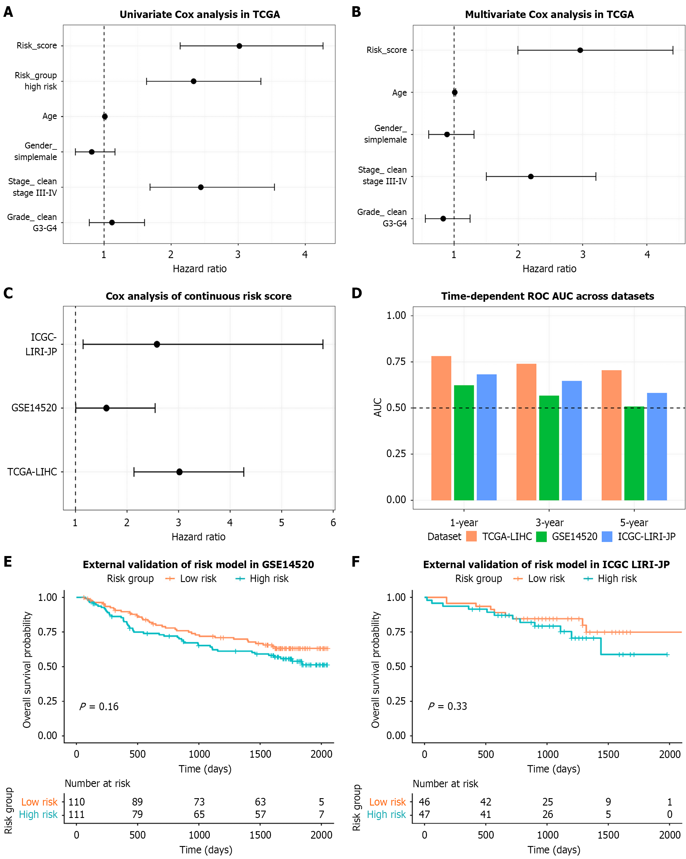
Figure 5 External performance evaluation and prognostic assessment of the six-gene risk score.
A: Univariate Cox regression analysis in The Cancer Genome Atlas liver hepatocellular carcinoma; B: Multivariable Cox regression analysis adjusted for age, sex, pathological stage, and tumor grade; C: Cox analysis of the continuous risk score across The Cancer Genome Atlas liver hepatocellular carcinoma, GSE14520, and ICGC-LIRI-JP; D: Summary of time-dependent areas under the curve across datasets; E: Kaplan-Meier curve in GSE14520; F: Kaplan-Meier curve in ICGC-LIRI-JP. TCGA-LIHC: The Cancer Genome Atlas liver hepatocellular carcinoma; AUC: Area under the curve; ROC: Receiver operating characteristic; ICGC-LIRI-JP: International Cancer Genome Consortium Liver Cancer-RIKEN Japan.
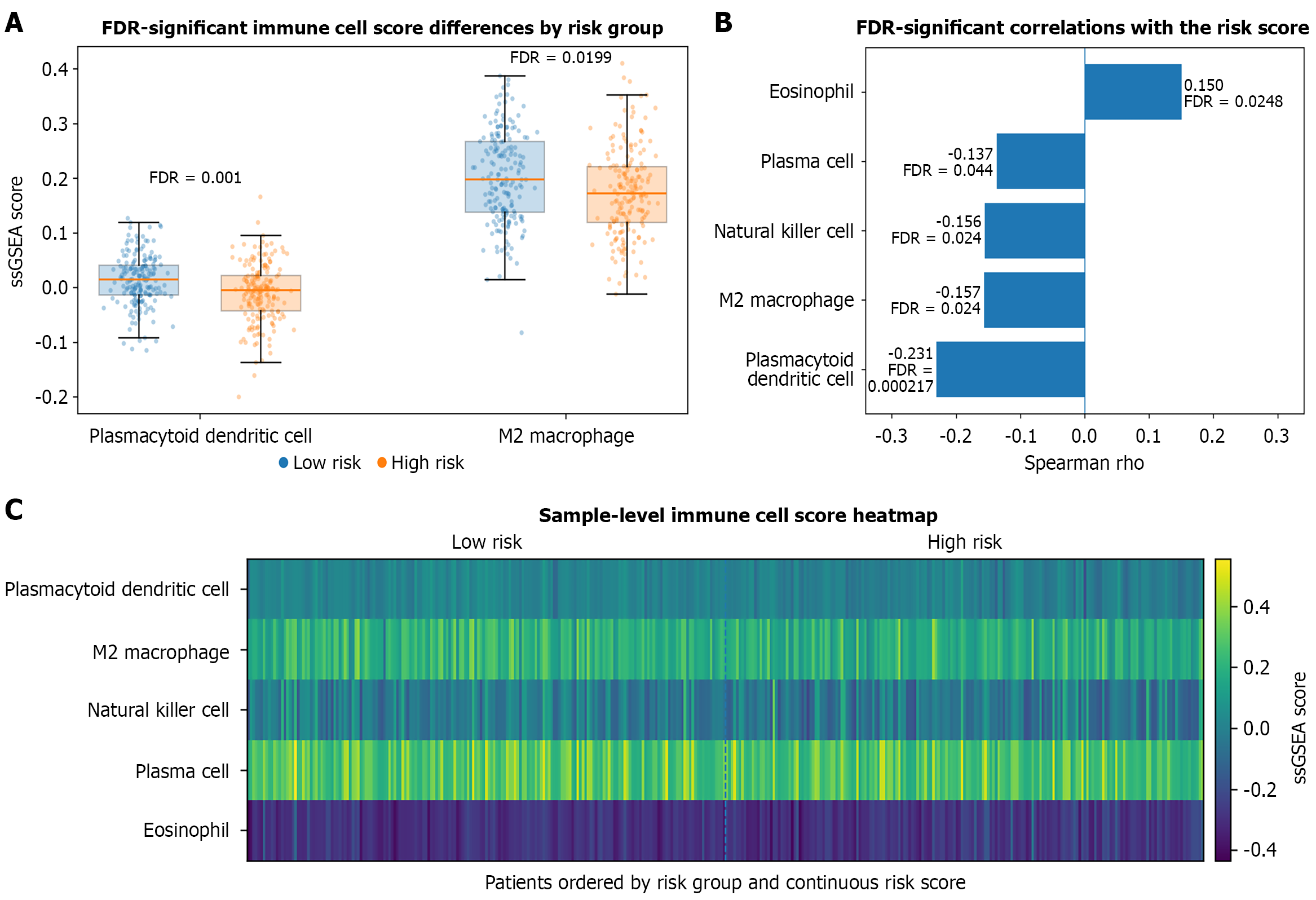
Figure 6 Single-sample gene set enrichment analysis-derived immune cell score patterns associated with the risk score.
A: Boxplots with individual patient observations for the two immune cell scores that differed significantly between the high- and low-risk groups after Benjamini-Hochberg correction; B: Spearman correlations between the continuous risk score and immune cell scores that remained significant after false discovery rate correction; C: Sample-level heatmap of the five immune cell scores that remained significant after false discovery rate correction, ordered by risk group and continuous risk score. FDR: False discovery rate; ssGSEA: Single-sample gene set enrichment analysis.
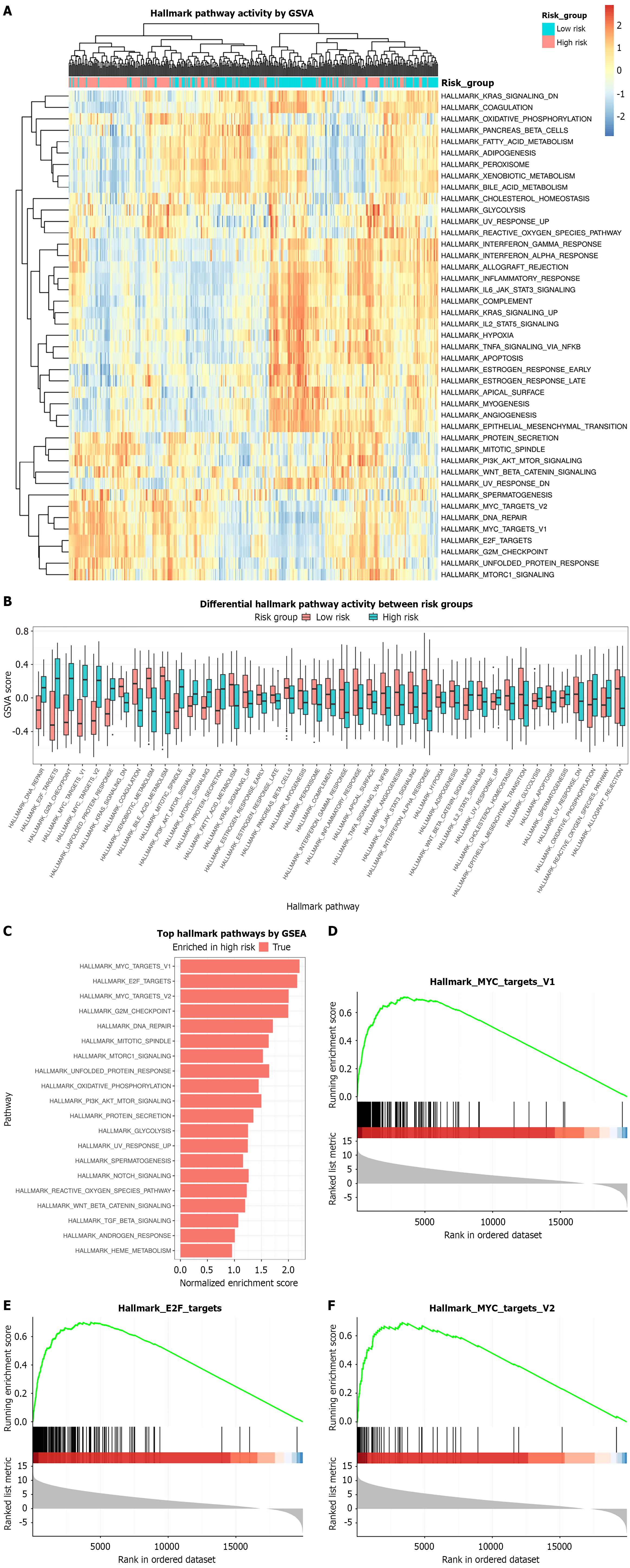
Figure 7 Hallmark pathway enrichment analysis of the ferroptosis-mitochondria-related risk signature.
A: Gene set variation analysis heatmap of differentially enriched Hallmark pathways; B: Boxplots of representative Hallmark pathways; C: Bar plot of normalized enrichment scores from gene set enrichment analysis; D-F: Gene set enrichment analysis plots for MYC targets V1, E2F targets, and MYC targets V2. GSEA: Gene set enrichment analysis; GSVA: Gene set variation analysis.
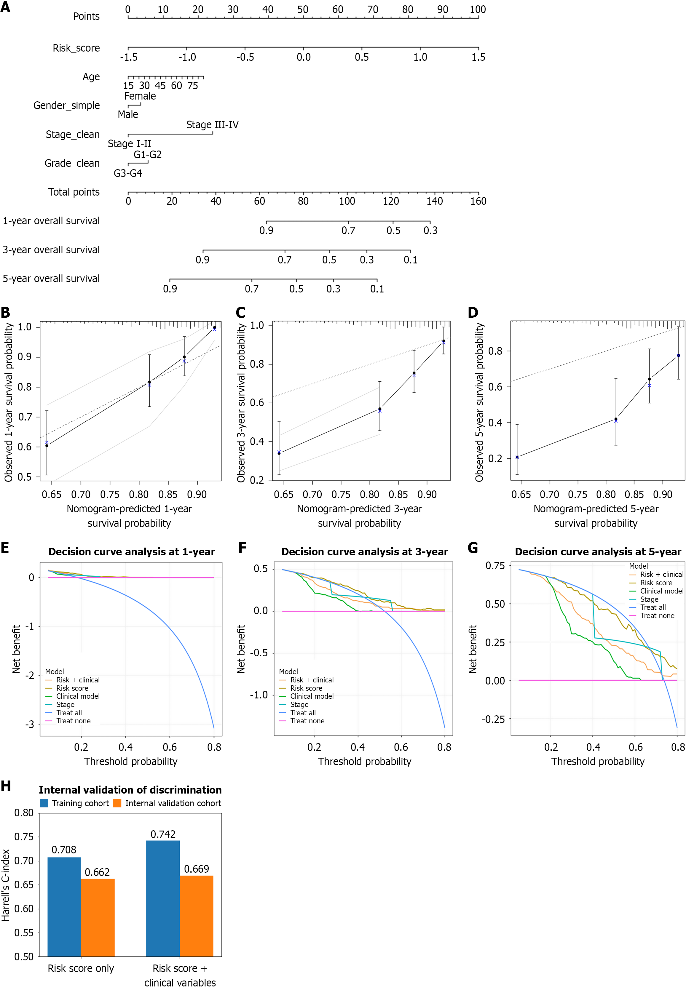
Figure 8 Nomogram construction and validation for the six-gene risk score in The Cancer Genome Atlas liver hepatocellular carcinoma.
A: Single multivariable Cox nomogram integrating risk score, age, sex, pathological stage, and tumor grade, with separate scales for 1-, 3-, and 5-year overall survival probabilities; B-D: Bootstrap calibration curves for 1-, 3-, and 5-year overall survival prediction; E-G: Decision curve analyses at 1, 3, and 5 years; H: Harrell’s C-index for the risk score-only and combined models in the training and internal validation cohorts.
- Citation: Han S, Feng ZW, Ran Y, Ma GP, Zhou YC, Lv JZ, Li XB, Wang CH, Wang LZ, Hu SP. Ferroptosis-mitochondria-related molecular subtypes and prognostic signature reveal immune and pathway heterogeneity in hepatocellular carcinoma. World J Gastrointest Oncol 2026; 18(11): 124061
- URL: https://www.wjgnet.com/1948-5204/full/v18/i11/124061.htm
- DOI: https://dx.doi.org/10.4251/wjgo.124061