
















Copyright: ©Author(s) 2026.
World J Stem Cells. Sep 26, 2026; 18(9): 124540
Published online Sep 26, 2026. doi: 10.4252/wjsc.124540
Published online Sep 26, 2026. doi: 10.4252/wjsc.124540
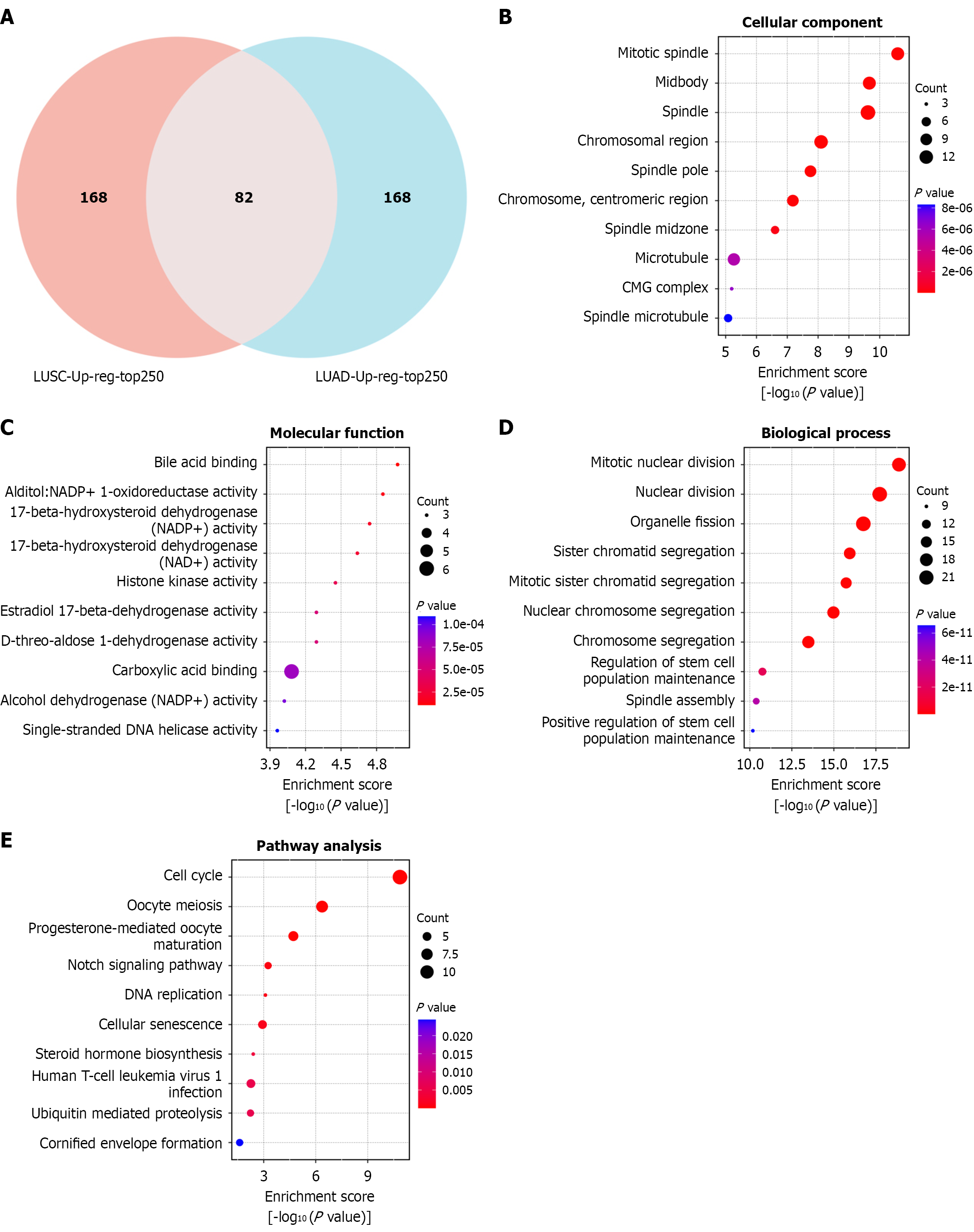
Figure 1 Enrichment analysis of mediator complex subunit 12-associated genes was performed based on The Cancer Genome Atlas database.
A: Venn diagram showing the overlapping genes positively correlated with mediator complex subunit 12 expression in lung adenocarcinoma and lung squamous cell carcinoma; B-D: Bubble plot of Gene Ontology enrichment analysis of the overlapping genes, including cellular component (B), molecular function (C), and biological process (D) enrichment; E: Bubble plot of Kyoto Encyclopedia of Genes and Genomes enrichment analysis of the overlapping genes. LUSC: Lung squamous cell carcinoma; LUAD: Lung adenocarcinoma.
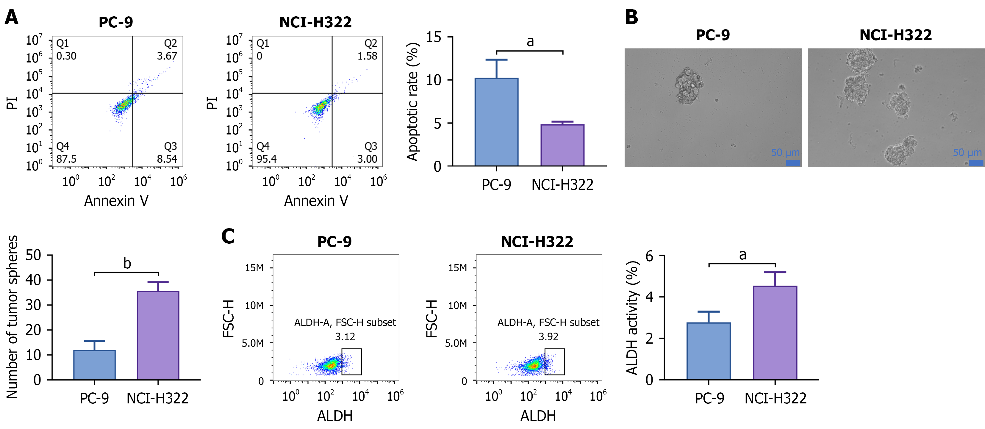
Figure 2 Mediator complex subunit 12 mutation was involved in the maintenance of cancer stem cells.
A: Flow cytometry was used to examine the apoptosis rates of NCI-H322 and PC9 cells; B: Sphere formation assay was used to evaluate the sphere-forming ability of NCI-H322 and PC9 cells; C: Flow cytometry was used to analyze the percentage of ALDH1-positive cells in NCI-H322 and PC9 cells. Data are presented as mean ± SD. aP < 0.05, bP < 0.01. All experiments were repeated 3 times.
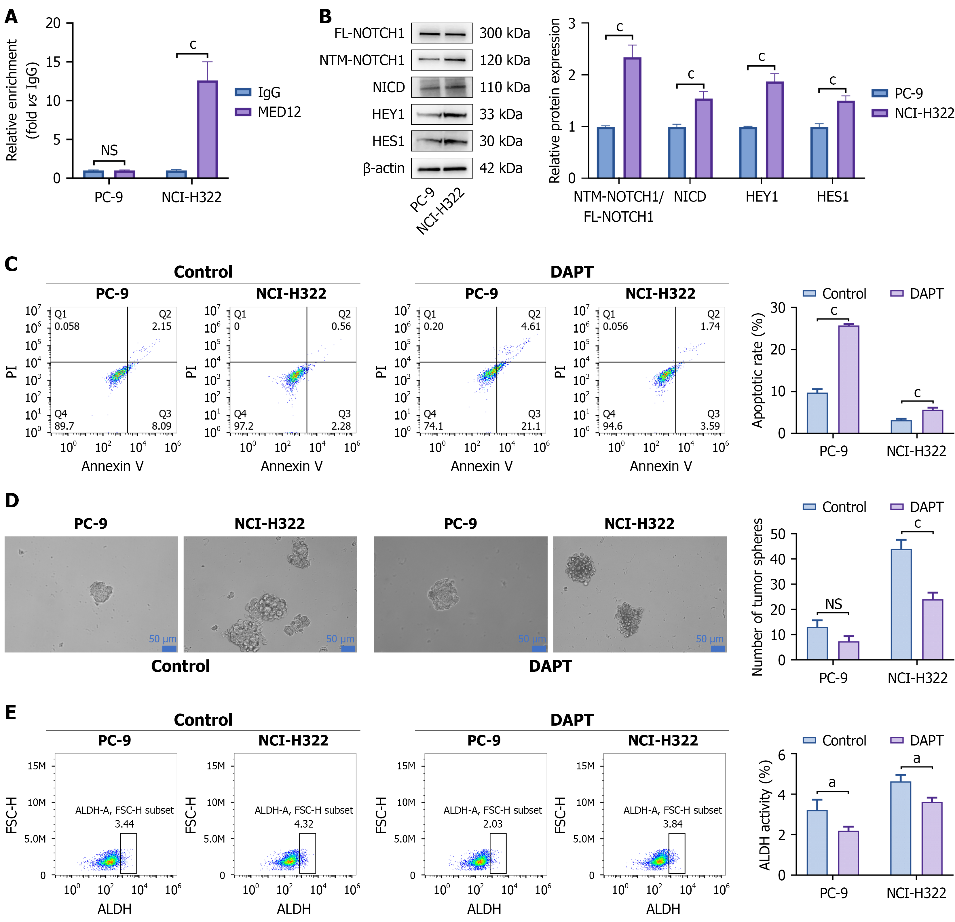
Figure 3 Mediator complex subunit 12 mutation maintained cancer stem cell properties via the Notch signaling pathway.
A: Chromatin immunoprecipitation-quantitative polymerase chain reaction analysis of mediator complex subunit 12 occupancy at the HES1 promoter in lung cancer cells; B: Western blot was performed to detect NOTCH1, N1ICD, HES1, and HEY1 protein expression in NCI-H322 and PC9 cells; C: Flow cytometry was used to examine the apoptosis rates of NCI-H322 and PC9 cells treated with or without DAPT; D: Sphere formation assay was used to evaluate the sphere-forming ability of NCI-H322 and PC9 cells treated with or without DAPT; E: Flow cytometry was used to analyze the percentage of ALDH1-positive cells in NCI-H322 and PC9 cells treated with or without DAPT. Data are presented as mean ± SD. All experiments were repeated 3 times. aP < 0.05, cP < 0.001. NS: Not significant; MED12: Mediator complex subunit 12; IgG: Immunoglobulin G.
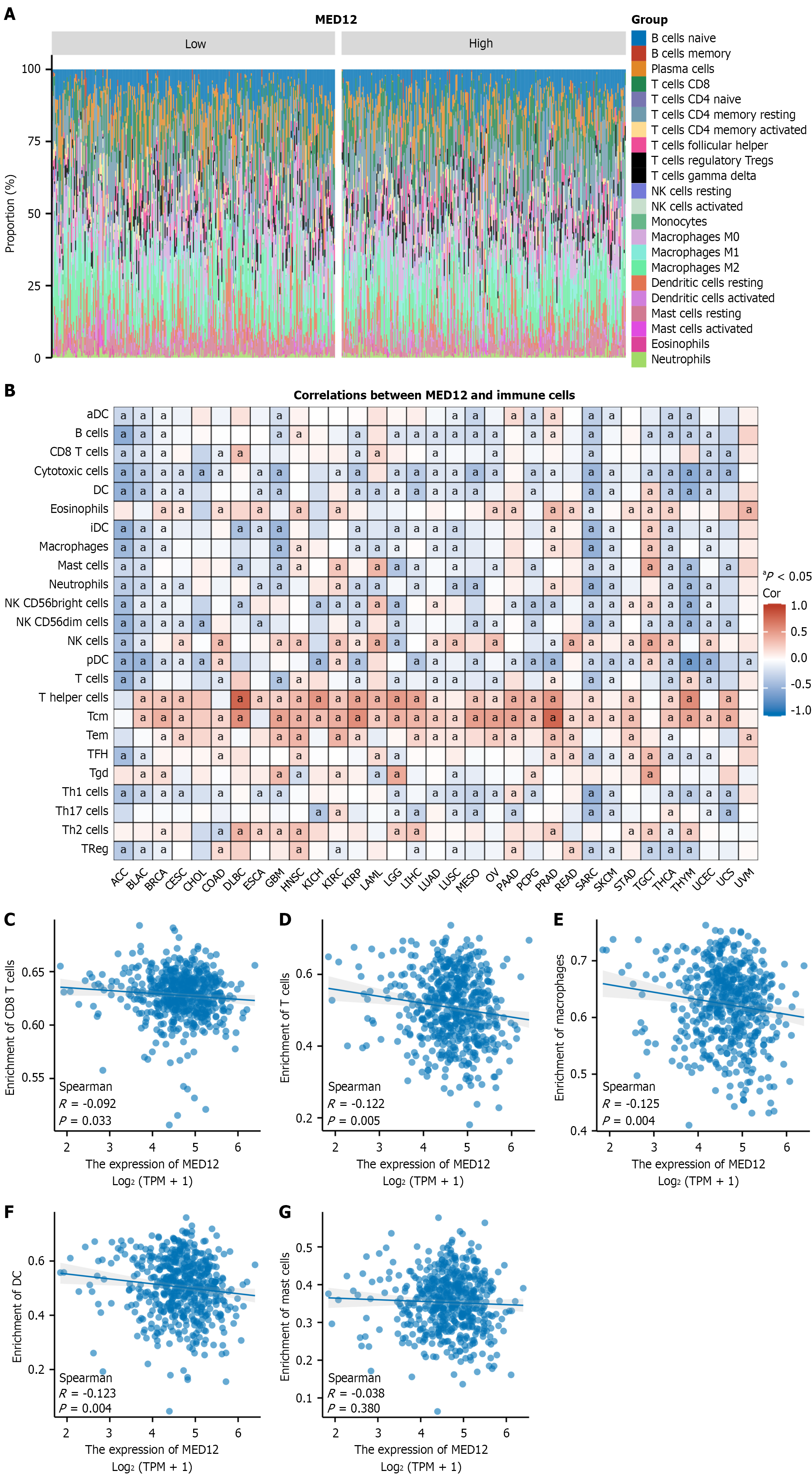
Figure 4 Analysis of the association between mediator complex subunit 12 expression and immune cell infiltration based on The Cancer Genome Atlas transcriptomic data.
A: Bar plot showing immune cell abundances in lung cancer samples with high and low mediator complex subunit 12 (MED12) expression; B: Correlation between MED12 expression and immune cell enrichment across pan-cancer; C-G: Scatter plots showing the correlation of MED12 expression with CD8+ T cells (C), T cells (D), macrophages (E), dendritic cells (F), and mast cells (G) in lung cancer patients. aP < 0.05. MED12: Mediator complex subunit 12; DC: Dendritic cell.
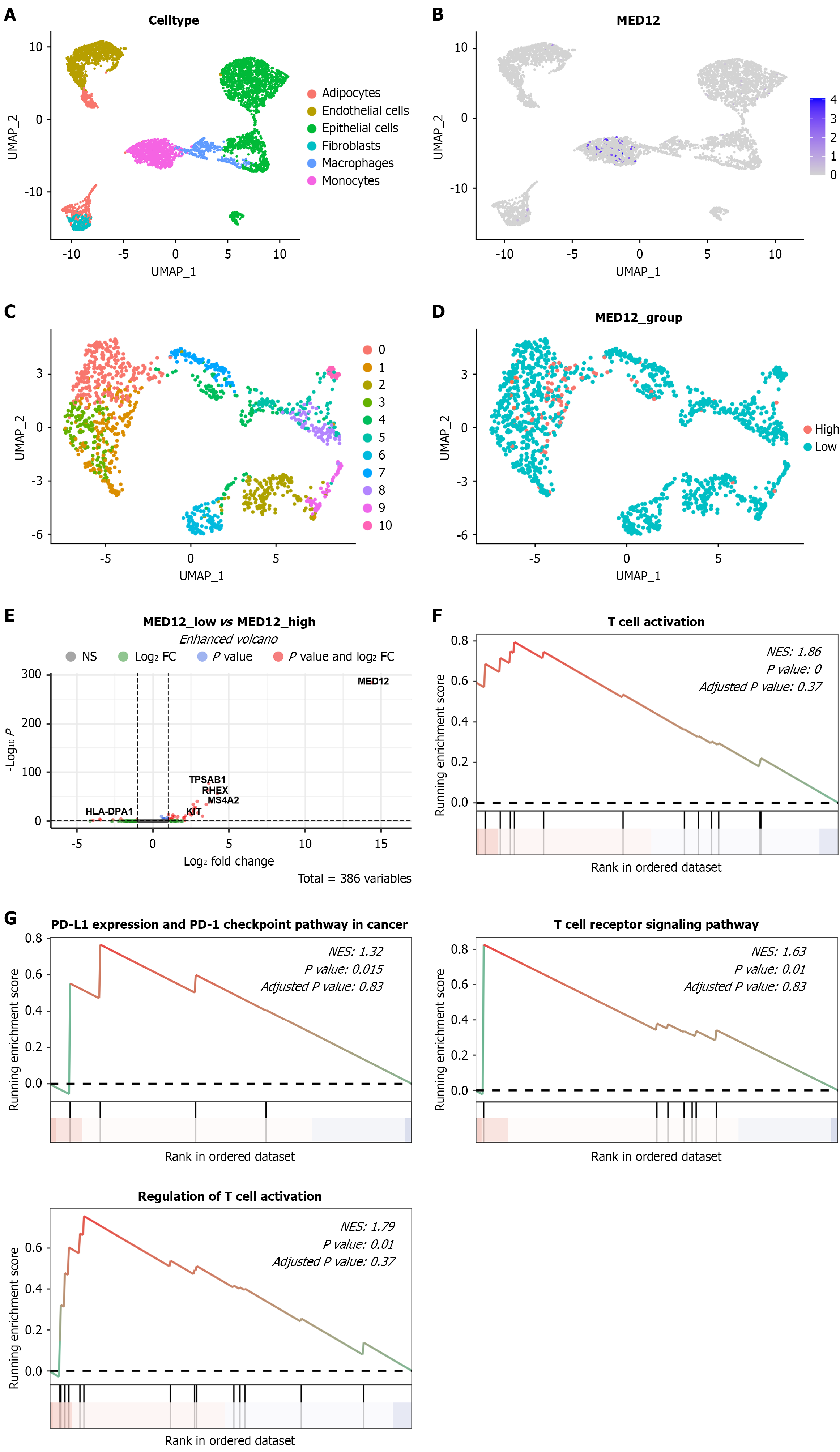
Figure 5 Elucidation of the association between mediator complex subunit 12 expression and immune cell infiltration based on single-cell sequencing data.
A: UMAP plot showing the identification of 6 cell subpopulations based on canonical cell markers; B: UMAP plot showing the distribution of mediator complex subunit 12 (MED12) expression across cell subpopulations; C: UMAP plot showing the sub clustering of macrophages and monocytes; D: UMAP plot showing the distribution of MED12-high and MED12-low expression groups within macrophage and monocyte subpopulations; E: Volcano plot showing differentially expressed genes between MED12-high and MED12-low cells; F: Gene set enrichment analysis (GSEA) enrichment analysis of differentially expressed genes based on the Gene Ontology database; G: GSEA enrichment analysis of differentially expressed genes based on the Kyoto Encyclopedia of Genes and Genomes database. MED12: Mediator complex subunit 12; PD-L1: Programmed death ligand-1.
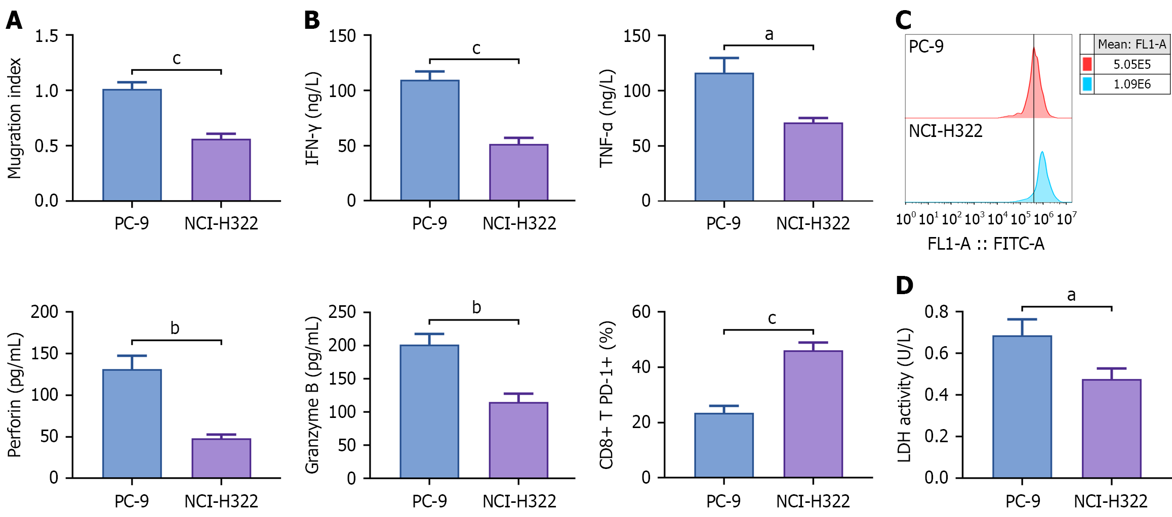
Figure 6 Mediator complex subunit 12 mutation mediated the inhibitory effect of cancer cells on the killing and chemotactic abilities of CD8+ T cells.
After co-culture of NCI-H322 and PC9 cells with CD8+ T cells, the following parameters of CD8+ T cells or tumor cells were analyzed. A: Transwell assay was performed to assess the migratory capacity of CD8+ T cells toward tumor cells; B: The expression of interferon-γ, tumor necrosis factor-α, perforin, and granzyme B in CD8+ T cells were measured by enzyme-linked immunosorbent assay; C: Flow cytometry was used to detect the percentage of programmed cell death 1-positive CD8+ T cells; D: The level of lactate dehydrogenase (LDH) in tumor cells was measured by LDH Cytotoxicity Assay Kit to evaluate the cytotoxic activity of CD8+ T cells. Data are presented as mean ± SD. aP < 0.05, bP < 0.01, cP < 0.001. All experiments were repeated 3 times. IFN: Interferon; TNF: Tumor necrosis factor.
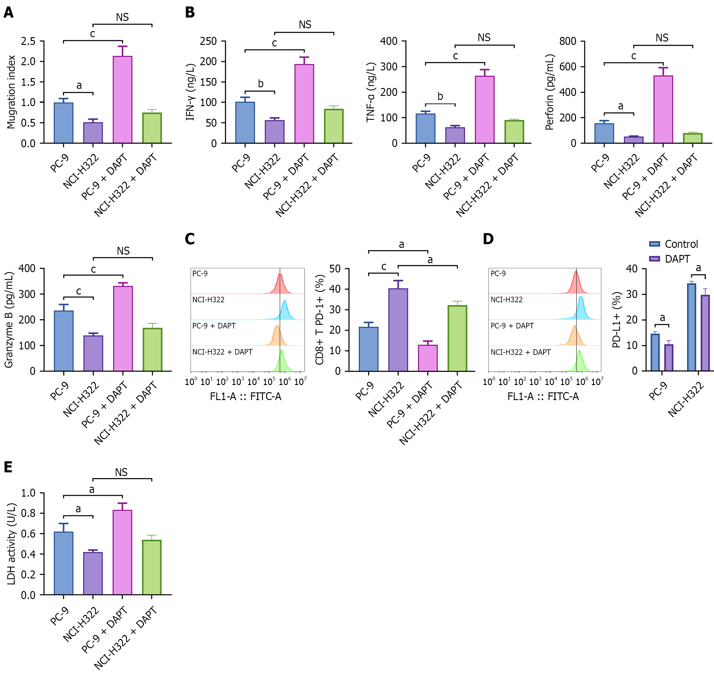
Figure 7 Mediator complex subunit 12 mutation mediated the inhibitory effect of cancer cells on CD8+ T cell killing and chemotaxis through activation of the Notch pathway.
After co-culturing CD8+ T cells with NCI-H322 and PC9 cells that had been treated with or without DAPT, the following parameters of the CD8+ T cells or tumor cells were analyzed. A: Transwell assay was performed to assess the migratory capacity of CD8+ T cells toward tumor cells; B: The expression of interferon-γ, tumor necrosis factor-α, perforin, and granzyme B in CD8+ T cells was measured by enzyme-linked immunosorbent assay; C: Flow cytometry was used to detect the percentage of programmed cell death 1-positive CD8+ T cells; D: Flow cytometry was used to detect the percentage of programmed death ligand-1-positive NCI-H322 and PC9 cells; E: The level of lactate dehydrogenase (LDH) in tumor cells was measured by LDH Cytotoxicity Assay Kit to evaluate the cytotoxic activity of CD8+ T cells. Data are presented as mean ± SD. All experiments were repeated 3 times. aP < 0.05, bP < 0.01, cP < 0.001. NS: Not significant; IFN: Interferon; TNF: Tumor necrosis factor.
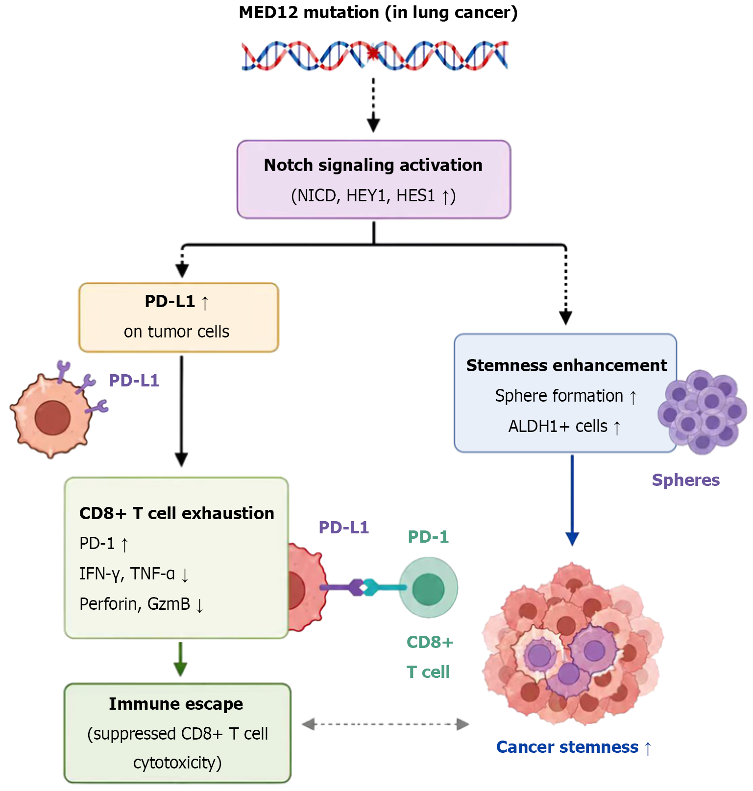
Figure 8 Graphic abstract.
Dashed arrows indicate functionally associated pathways observed in the NCI-H322 cell model. Definitive causal attribution of Notch activation to the mediator complex subunit 12 mutation requires further isogenic validation. MED12: Mediator complex subunit 12; PD-1: Programmed cell death 1; IFN: Interferon; TNF: Tumor necrosis factor; PD-L1: Programmed death ligand-1; GzmB: Granzyme B.
- Citation: Yang Y, Zhong JL, Liu JY. MED12 mutation activates Notch signaling to enhance cancer stemness and suppress CD8+ T cell cytotoxicity in lung cancer. World J Stem Cells 2026; 18(9): 124540
- URL: https://www.wjgnet.com/1948-0210/full/v18/i9/124540.htm
- DOI: https://dx.doi.org/10.4252/wjsc.124540