Published online Dec 26, 2022. doi: 10.12998/wjcc.v10.i36.13396
Peer-review started: September 5, 2022
First decision: November 11, 2022
Revised: November 16, 2022
Accepted: December 5, 2022
Article in press: December 5, 2022
Published online: December 26, 2022
Processing time: 108 Days and 14.1 Hours
Post-traumatic blunt pericardial injury is a rare condition with only a few re
In this case, we report a blunt pericardial injury undetected on preoperative tr
Post-traumatic blunt pericardial ruptures should be considered in patients with blunt chest trauma showing abnormal vital signs and ECG changes.
Core Tip: Post-traumatic blunt pericardial rupture is a rare but fatal injury that is usually diagnosed with computed tomography upon admission, but the condition can be asymptomatic or misdiagnosed. If a patient with blunt chest trauma shows fluctuating vital signs and accompanying electrocardiogram abnormalities, pericardial rupture and cardiac herniation should be considered.
- Citation: Yoon SY, Ye JB, Seok J. Undetected traumatic cardiac herniation like playing hide-and-seek-delayed incidental findings during surgical stabilization of flail chest: A case report. World J Clin Cases 2022; 10(36): 13396-13401
- URL: https://www.wjgnet.com/2307-8960/full/v10/i36/13396.htm
- DOI: https://dx.doi.org/10.12998/wjcc.v10.i36.13396
Post-traumatic blunt pericardial injury is rare but has occasionally been reported[1-4]. Once diagnosed, surgical repair is mandatory as this condition can trigger devastating complications[5,6]. However, in some cases, cardiac herniations may be misdiagnosed or masked by concomitant injury. In this case report, we share our experience with a blunt pericardial rupture with no specific symptoms apart from intermittent electrocardiogram (ECG) changes; the case was undiagnosed even after chest computed tomography (CT) and transthoracic echocardiography (TTE) in the preoperative period.
A 46-year-old male patient who was involved in a motor vehicle accident presented with a left abdominal wall injury and severe chest pain.
The patient had no specific history of present illness.
The patient had no specific history of past illness.
The patient had no personal or familial disease.
Tenderness and rebound tenderness were observed in the lower left quadrant of the abdomen. Crepitus and tenderness were present in the left chest wall, accompanied by decreased breath sounds. Symmetrical chest wall expansion during breathing was observed with no initial flail motion.
Initial arterial blood gas analysis showed a pH of 7.4, PO2 of 80.3 mmHg, and PCO2 of 34.0 mmHg on room air. Cardiac enzymes were elevated at troponin T 244 ng/L (normal range: < 14 ng/L), CK-MB 23.32 ng/L (normal range: < 4.94 ng/L), and CPK 4176 U/L (normal range: 58–348 U/L).
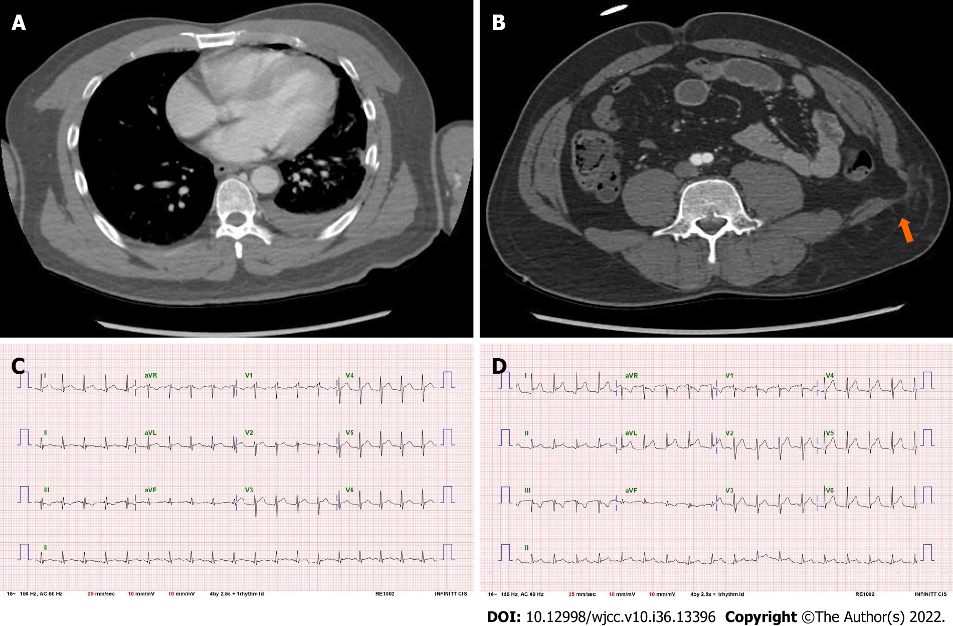
Chest CT revealed multiple rib fractures with hemopneumothorax in the left hemithorax. No abnormalities were observed in the mediastinum. Abdominal CT revealed a bowel herniation through an abdominal wall defect. The initial ECG showed an elevated ST segment in the anterior leads; however, it was deemed to be insignificant (Figure 1).
The patient was initially diagnosed with multiple rib fractures and hemopneumothorax in the left chest. Panperitonitis due to bowel perforation was suspected, and blunt cardiac contusion was considered highly likely.
A 24-French thoracic drainage catheter was placed, and the hemothorax was confirmed to be minor. Despite the perioperative risks associated with cardiac contusions, the patient underwent emergent surgery to rule out abdominal organ injuries. The peritoneum was intact, and no significant findings were observed except for several pelvic muscle tears.
Postoperative care in the surgical intensive care unit (SICU) was provided as usual. Blood tests for elevated cardiac enzymes were repeated every 4 h and showed gradual improvement. The patient did not complain of worsening chest pain, which was successfully controlled with analgesics. However, the patient’s systolic blood pressure fluctuated intermittently with prominent ECG changes. Although the changes were not the classical reciprocal findings observed for the myocardial infarction (MI), we still found it necessary to confirm the patient’s cardiac condition (Figure 1).
On Day 2 of hospitalization, TTE was performed to check for cardiac conditions. The patient was confirmed to have neither pericardial fluid collection nor abnormal cardiac movements. The patient’s ECG remained variable, fluctuating from normal to prominent ST elevations, and we regarded these findings as non-meaningful and indicative of the usual transient changes after cardiac contusion.
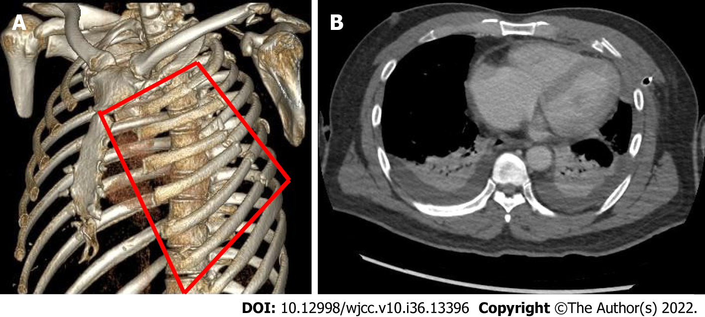
On Day 3 of hospitalization, the patient complained of dyspnea and difficulty with expectoration. Paradoxical asymmetric chest wall motion was newly observed on the left anterior chest wall to the extent where the chest wall moved in rhythm with the patient’s heartbeat. Despite proper support, including higher oxygen supplementation via a high-flow nasal cannula, the patient’s condition gradually deteriorated. Follow-up chest CT revealed a worsening lung condition. However, the mediastinum was relatively clear (Figure 2). After thoroughly discussing the risks associated with cardiac contusions with anesthesiologists, we decided to perform surgical stabilization of rib fractures (SSRF).
A flail chest due to multiple rib fractures and blunt cardiac contusion were considered as additionally diagnosed conditions.
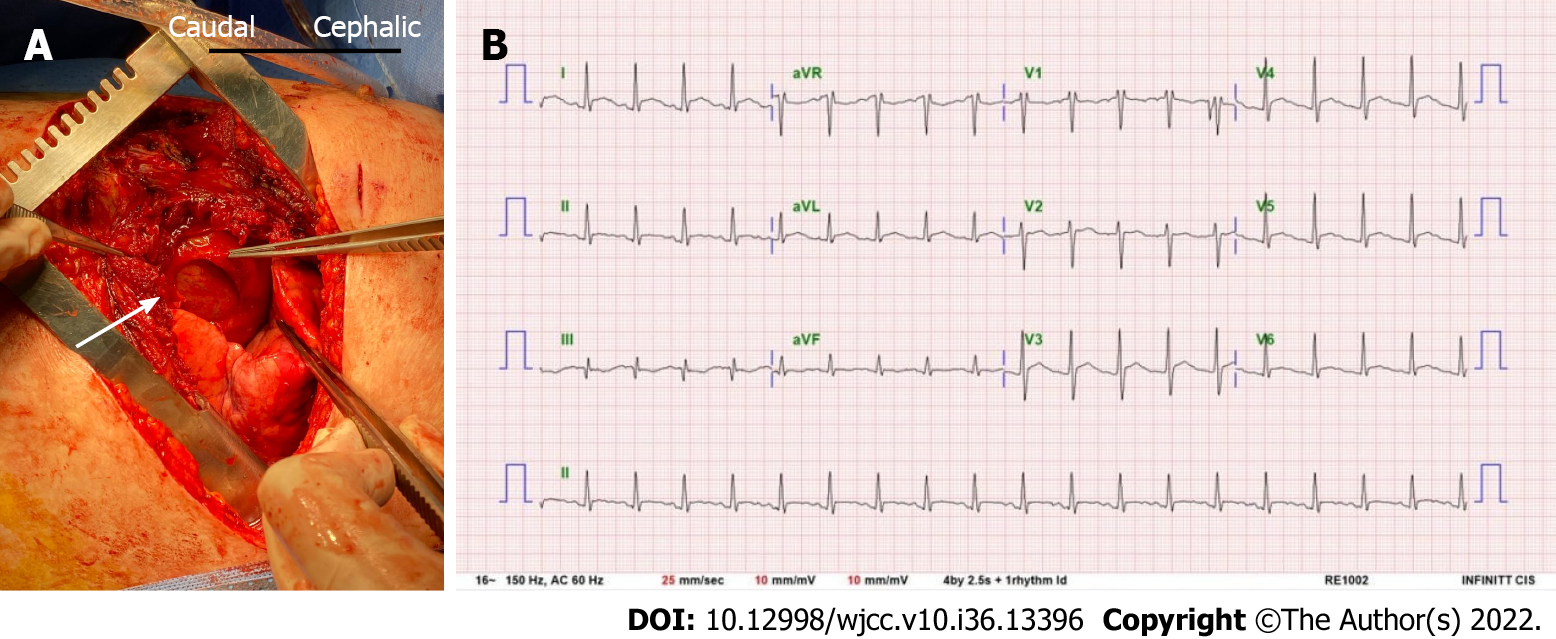
The patient underwent SSRF on Day four of hospitalization. The surgery was performed routinely under general anesthesia with a single endotracheal intubation, with the patient placed in the 45-degree semi-lateral right decubitus position. An anterolateral thoracotomy was performed in the fifth intercostal space from the median border of the left pectoralis major muscle to the mid-axillary line. After opening the fifth intercostal space, a large hole was identified in the pericardium (Figure 3). The inferolateral left ventricular wall was visible through the hole and was covered by a thin whitish fibrin clot. We explored the pericardial space with an index finger and confirmed no apparent abnormalities.
Post-traumatic pericardial rupture with a flail chest by multiple rib fractures.
The pericardium was subsequently repaired with 4-0 Prolene sutures. After stabilizing the fractured ribs with plates and screws and closing the wound, the patient was transferred to the SICU in stable condition.
The patient was successfully weaned off the mechanical ventilator two days following the operation (Day 5 of hospitalization). The postoperative ECG findings were still abnormal but had improved (Figure 3), and the patient was transferred to the general ward on Day 7 of hospitalization.
In most cases, pericardial ruptures are diagnosed or suspected on chest CT on admission. Pneumo- or hemopericardium strongly suggests heart damage; therefore, these are considered strong indicators that intensive treatment, such as surgery, will be required. However, in the classification of traumatic cardiac dislocations proposed by Graef et al[3], grade I pericardial rupture without dislocation of the heart can barely be detected on CT scans, and entirely secondary findings in patients with a diaphragm with intrathoracic herniation of abdominal organs can be diagnosed by chest radiography or CT[7]. Similarly, in our case, no preoperative examination suggested pericardial rupture. In cases of silent pericardial rupture accompanied by intermittent symptomatic cardiac herniation, as in our patient, it was almost impossible to suspect pericardial rupture until a thoracotomy was performed. If the patient had not worsened due to a flail chest, the probability of finding pericardial rupture would have been extremely low.
Motto et al[8] reported that cardiac herniation could be intermittent; therefore, sequential evaluation is recommended. We performed serial laboratory follow-ups for elevated cardiac enzyme levels, consecutive chest CTs, and preoperative TTE. In our case, however, the pericardial hole was only 4 cm × 4 cm and was too narrow to cause a severe symptomatic cardiac herniation. Instead, we hypothesized that it may have caused intermittent ECG changes and vital instability, resembling MI. In addition, pain from the patient’s rib fractures may have masked chest discomfort due to cardiac herniation. Many patients with cardiac contusions show elevated cardiac enzyme levels and abnormal ECG findings[9]. Finally, we deemed these findings to be typical of cardiac contusions, which do not require treatment. However, we believe that this case may not be unique, and such a mistake is common in practice.
Post-traumatic blunt pericardial rupture is a rare but fatal injury that is usually diagnosed by CT upon admission but may be asymptomatic and misdiagnosed. If a patient with blunt chest trauma shows fluctuating vital signs and accompanying ECG abnormalities, pericardial rupture and cardiac herniation should be considered.
Provenance and peer review: Unsolicited article; Externally peer reviewed.
Peer-review model: Single blind
Specialty type: Medicine, research and experimental
Country/Territory of origin: South Korea
Peer-review report’s scientific quality classification
Grade A (Excellent): 0
Grade B (Very good): B
Grade C (Good): C
Grade D (Fair): 0
Grade E (Poor): 0
P-Reviewer: Li Q, China; Ong H, Malaysia S-Editor: Fan JR L-Editor: A P-Editor: Fan JR
| 1. | Cook F, Mounier R, Martin M, Dhonneur G. Late diagnosis of post-traumatic ruptured pericardium with cardiac herniation. Can J Anaesth. 2017;64:94-95. [RCA] [PubMed] [DOI] [Full Text] [Cited by in Crossref: 3] [Cited by in RCA: 2] [Article Influence: 0.2] [Reference Citation Analysis (0)] |
| 2. | Gorospe L, Almeida-Aróstegui NA, Miguelena-Hycka J. Delayed Incidental Diagnosis of Posttraumatic Cardiac Herniation. Rev Esp Cardiol (Engl Ed). 2019;72:343. [RCA] [PubMed] [DOI] [Full Text] [Cited by in Crossref: 1] [Cited by in RCA: 1] [Article Influence: 0.2] [Reference Citation Analysis (0)] |
| 3. | Graef F, Walter S, Baur A, Tsitsilonis S, Moroder P, Kempfert J, Märdian S. Traumatic cardiac dislocation-A case report and review of the literature including a new classification system. J Trauma Acute Care Surg. 2019;87:944-953. [RCA] [PubMed] [DOI] [Full Text] [Cited by in Crossref: 3] [Cited by in RCA: 7] [Article Influence: 1.4] [Reference Citation Analysis (0)] |
| 4. | Lajmi M, Ben Ismail I, Ragmoun W, Messaoudi H, Lahdhili H, Chenik S. Post traumatic left cardiac luxation: A case report. Int J Surg Case Rep. 2020;71:205-208. [RCA] [PubMed] [DOI] [Full Text] [Full Text (PDF)] [Reference Citation Analysis (0)] |
| 5. | Kamiyoshihara M, Igai H, Kawatani N, Ibe T. Right or Left Traumatic Pericardial Rupture: Report of a Thought-Provoking Case. Ann Thorac Cardiovasc Surg. 2016;22:49-51. [RCA] [PubMed] [DOI] [Full Text] [Cited by in Crossref: 4] [Cited by in RCA: 5] [Article Influence: 0.5] [Reference Citation Analysis (0)] |
| 6. | Guenther T, Rinderknecht T, Phan H, Wozniak C, Rodriquez V. Pericardial rupture leading to cardiac herniation after blunt trauma. Trauma Case Rep. 2020;27:100309. [RCA] [PubMed] [DOI] [Full Text] [Full Text (PDF)] [Cited by in Crossref: 1] [Cited by in RCA: 3] [Article Influence: 0.6] [Reference Citation Analysis (0)] |
| 7. | Fulda G, Brathwaite CE, Rodriguez A, Turney SZ, Dunham CM, Cowley RA. Blunt traumatic rupture of the heart and pericardium: a ten-year experience (1979-1989). J Trauma. 1991;31:167-72; discussion 172. [RCA] [PubMed] [DOI] [Full Text] [Cited by in Crossref: 188] [Cited by in RCA: 173] [Article Influence: 5.1] [Reference Citation Analysis (0)] |
| 8. | Motto DA, Kurapati S, Atencio DC, Miller MA, Stahlfeld KR. Pericardial Rupture with Intermittent Cardiac Luxation. Tex Heart Inst J. 2018;45:115-116. [RCA] [PubMed] [DOI] [Full Text] [Cited by in Crossref: 3] [Cited by in RCA: 6] [Article Influence: 0.9] [Reference Citation Analysis (0)] |
| 9. | Alborzi Z, Zangouri V, Paydar S, Ghahramani Z, Shafa M, Ziaeian B, Radpey MR, Amirian A, Khodaei S. Diagnosing Myocardial Contusion after Blunt Chest Trauma. J Tehran Heart Cent. 2016;11:49-54. [PubMed] |


