Copyright
©The Author(s) 2022.
World J Clin Cases. Dec 26, 2022; 10(36): 13396-13401
Published online Dec 26, 2022. doi: 10.12998/wjcc.v10.i36.13396
Published online Dec 26, 2022. doi: 10.12998/wjcc.v10.i36.13396
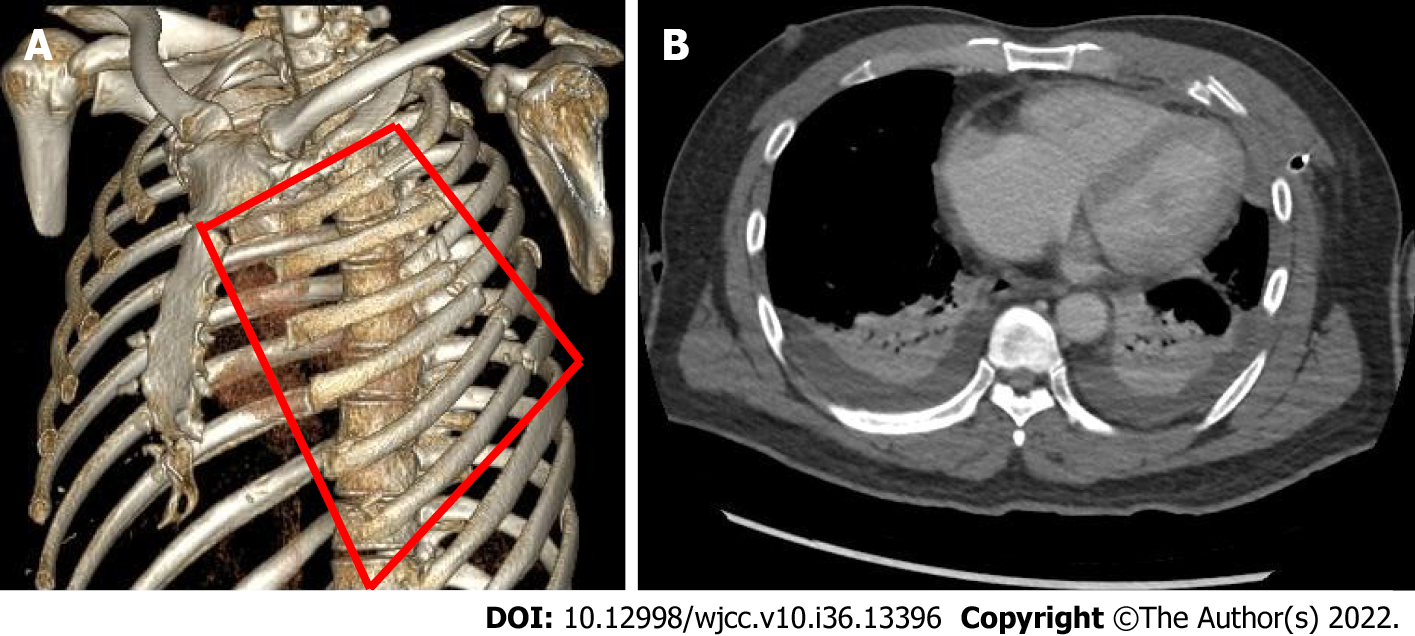
Figure 2 3-dimensional contrast chest computed tomography.
A: A 3-dimensional (3D) contrast chest computed tomography (CT) with rib rendering. The anterolateral flail segment (indicated by the red lines) was located on the left rib cage; B: An axial cut of the 3D chest CT showing bilateral pleural effusion with collapsed lungs, with no evidence of heart injury.
- Citation: Yoon SY, Ye JB, Seok J. Undetected traumatic cardiac herniation like playing hide-and-seek-delayed incidental findings during surgical stabilization of flail chest: A case report. World J Clin Cases 2022; 10(36): 13396-13401
- URL: https://www.wjgnet.com/2307-8960/full/v10/i36/13396.htm
- DOI: https://dx.doi.org/10.12998/wjcc.v10.i36.13396


