Copyright
©The Author(s) 2022.
World J Clin Cases. Jan 21, 2022; 10(3): 1093-1098
Published online Jan 21, 2022. doi: 10.12998/wjcc.v10.i3.1093
Published online Jan 21, 2022. doi: 10.12998/wjcc.v10.i3.1093
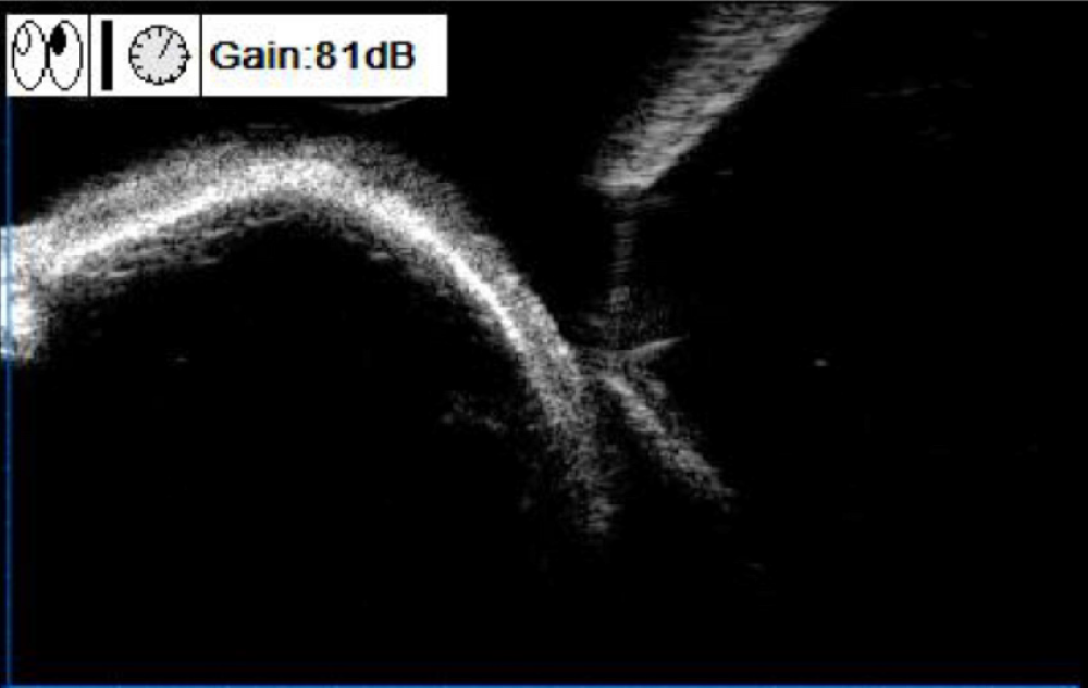
Figure 2 Preoperative ultrasound biomicroscopy image of the mass in the superior temporal quadrant of the left eye (case 1).
A strong oval echo was observed in the superficial sclera under the bulbar conjunctiva, with a clear boundary obscuring the lower echo.
- Citation: Wang YC, Wang ZZ, You DB, Wang W. Epibulbar osseous choristoma: Two case reports. World J Clin Cases 2022; 10(3): 1093-1098
- URL: https://www.wjgnet.com/2307-8960/full/v10/i3/1093.htm
- DOI: https://dx.doi.org/10.12998/wjcc.v10.i3.1093


