Copyright
©The Author(s) 2021.
World J Clin Cases. Jun 26, 2021; 9(18): 4810-4816
Published online Jun 26, 2021. doi: 10.12998/wjcc.v9.i18.4810
Published online Jun 26, 2021. doi: 10.12998/wjcc.v9.i18.4810
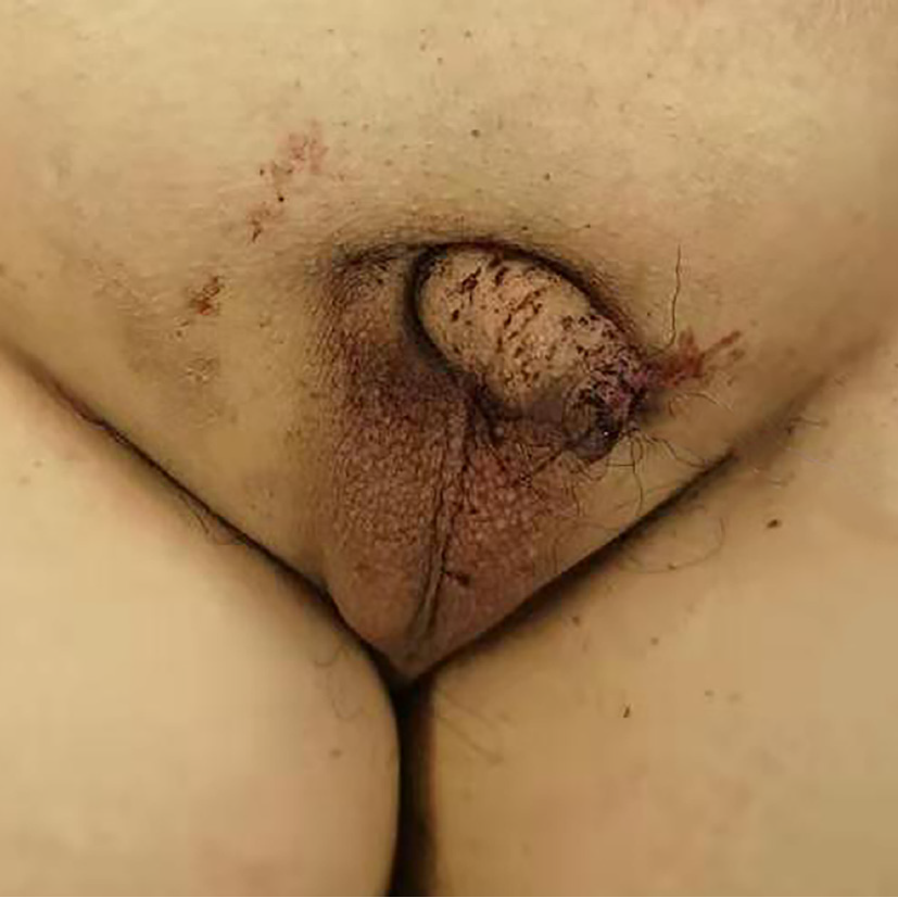
Figure 1 Physical examination showed that the penis and scrotum were underdeveloped, and the testis was impalpable in the scrotum.
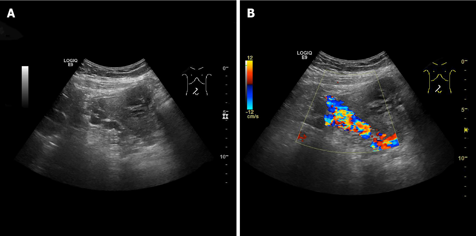
Figure 2 Color Doppler ultrasound images.
A: Color Doppler ultrasound demonstrated many twisted tube-like echoes around the bladder; B: Color Doppler flow imaging showed color flow signal inside the tube-like echoes.
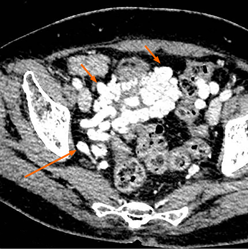
Figure 3 Axial contrast-enhanced computed tomography image demonstrating multiple tortuous and thickened veins on the anterior wall and both sidewalls of the bladder (short arrow).
The dilated vesical varices on the right side drained into the internal iliac vein (long arrow).
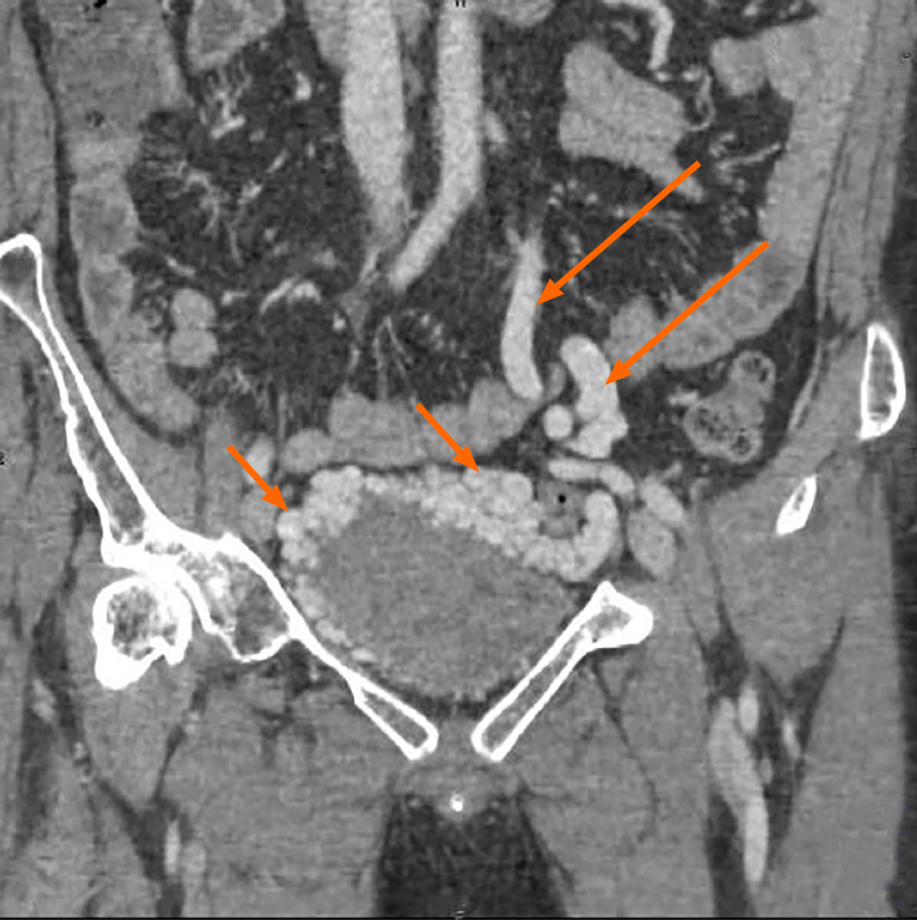
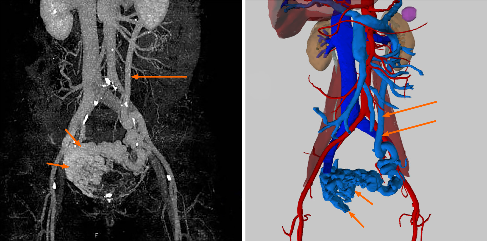
Figure 4 Contrast-enhanced coronal computed tomography-reconstructed images demonstrating abnormally dilated blood vessels (short arrow) surrounding the bladder, and the enlargement of inferior mesenteric veins (long arrow).
Figure 5 Three-dimensional visualization technology clearly showed that there were many abnormally dilated blood vessels surrounding the bladder in the pelvis (short arrow).
In this patient, the dilated vesical varices on the right side drained into the internal iliac vein and on the left side was connected with the inferior mesenteric vein (long arrow), and finally entered into the splenic vein.
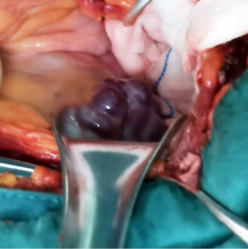
Figure 6 A large number of tortuous and swollen veins were seen surrounding the urinary bladder, twisting each other into a mass.
- Citation: Wei ZJ, Zhu X, Yu HT, Liang ZJ, Gou X, Chen Y. Severe hematuria due to vesical varices in a patient with portal hypertension: A case report. World J Clin Cases 2021; 9(18): 4810-4816
- URL: https://www.wjgnet.com/2307-8960/full/v9/i18/4810.htm
- DOI: https://dx.doi.org/10.12998/wjcc.v9.i18.4810


