Copyright
©The Author(s) 2021.
World J Clin Cases. Jun 26, 2021; 9(18): 4810-4816
Published online Jun 26, 2021. doi: 10.12998/wjcc.v9.i18.4810
Published online Jun 26, 2021. doi: 10.12998/wjcc.v9.i18.4810
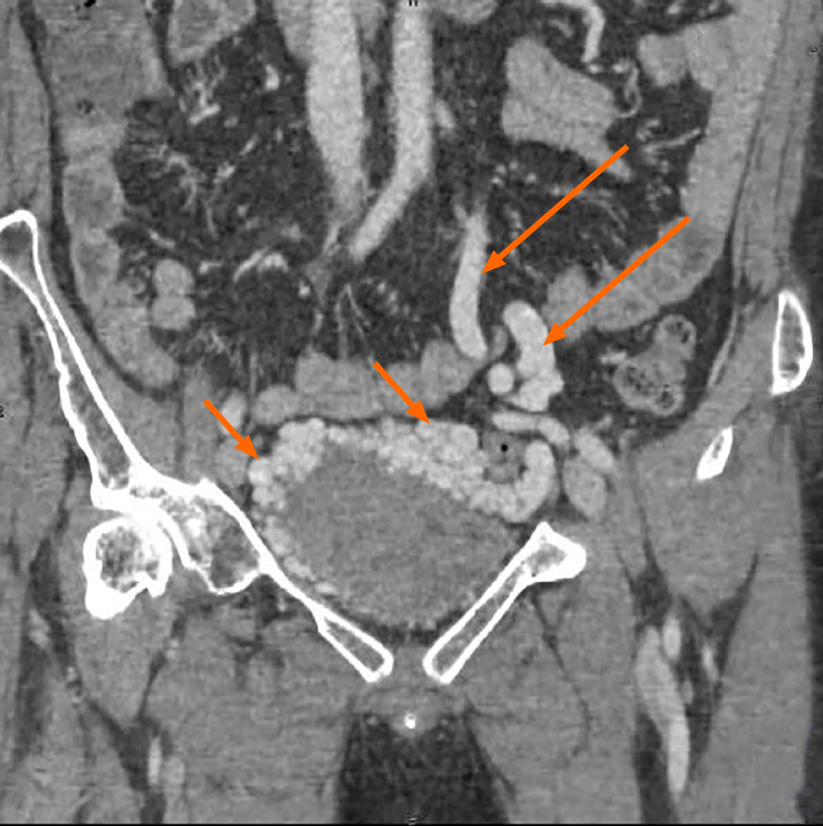
Figure 4 Contrast-enhanced coronal computed tomography-reconstructed images demonstrating abnormally dilated blood vessels (short arrow) surrounding the bladder, and the enlargement of inferior mesenteric veins (long arrow).
- Citation: Wei ZJ, Zhu X, Yu HT, Liang ZJ, Gou X, Chen Y. Severe hematuria due to vesical varices in a patient with portal hypertension: A case report. World J Clin Cases 2021; 9(18): 4810-4816
- URL: https://www.wjgnet.com/2307-8960/full/v9/i18/4810.htm
- DOI: https://dx.doi.org/10.12998/wjcc.v9.i18.4810


