Copyright
©The Author(s) 2021.
World J Clin Cases. Jun 26, 2021; 9(18): 4810-4816
Published online Jun 26, 2021. doi: 10.12998/wjcc.v9.i18.4810
Published online Jun 26, 2021. doi: 10.12998/wjcc.v9.i18.4810
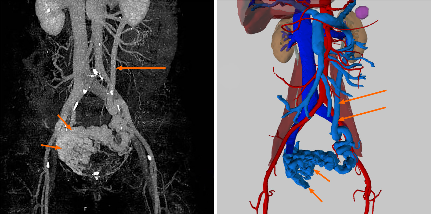
Figure 5 Three-dimensional visualization technology clearly showed that there were many abnormally dilated blood vessels surrounding the bladder in the pelvis (short arrow).
In this patient, the dilated vesical varices on the right side drained into the internal iliac vein and on the left side was connected with the inferior mesenteric vein (long arrow), and finally entered into the splenic vein.
- Citation: Wei ZJ, Zhu X, Yu HT, Liang ZJ, Gou X, Chen Y. Severe hematuria due to vesical varices in a patient with portal hypertension: A case report. World J Clin Cases 2021; 9(18): 4810-4816
- URL: https://www.wjgnet.com/2307-8960/full/v9/i18/4810.htm
- DOI: https://dx.doi.org/10.12998/wjcc.v9.i18.4810


