Copyright
©The Author(s) 2025.
World J Clin Cases. Aug 26, 2025; 13(24): 107555
Published online Aug 26, 2025. doi: 10.12998/wjcc.v13.i24.107555
Published online Aug 26, 2025. doi: 10.12998/wjcc.v13.i24.107555
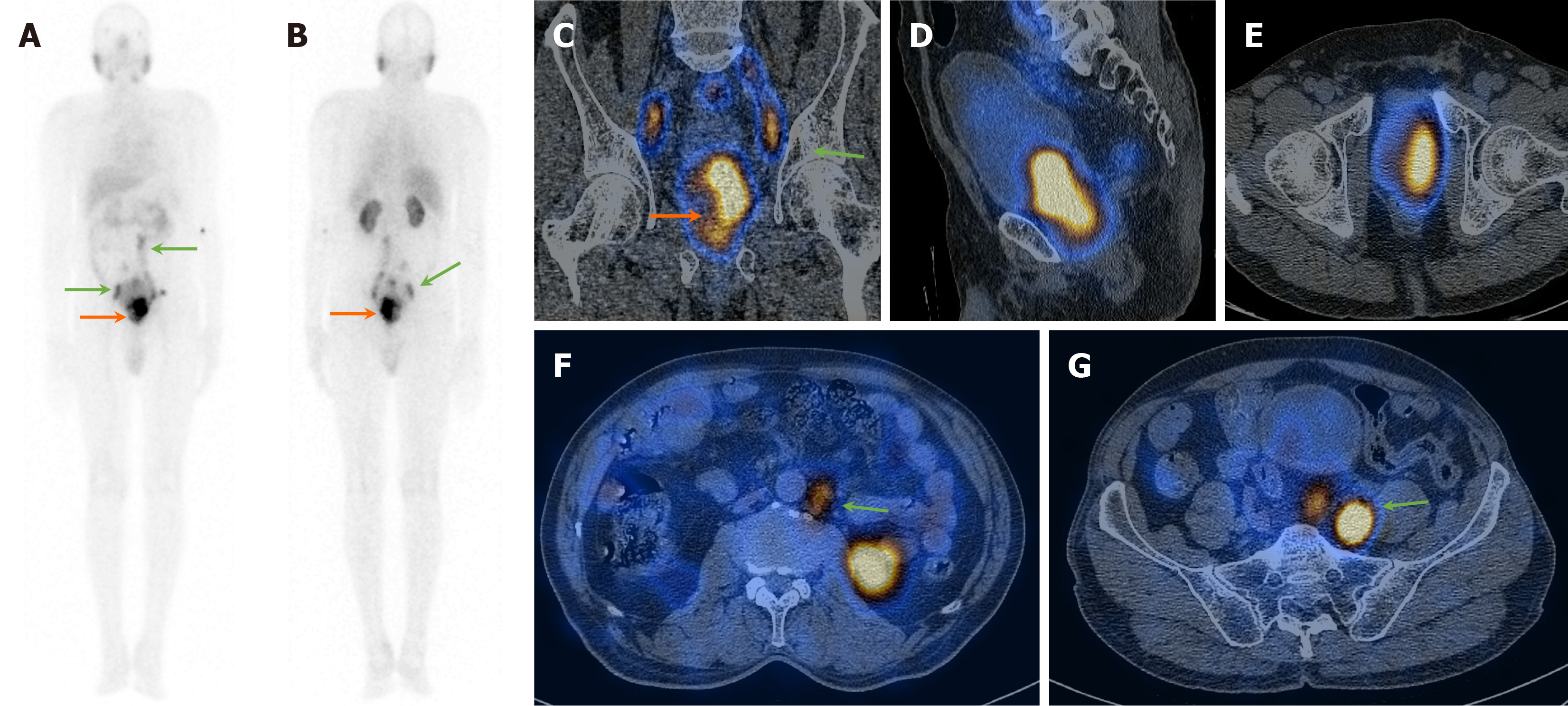
Figure 1 A 59-year-old man presented with hematuria and difficulty urination, S.
PSA > 100 ng/dL, and a suspicious lesion in the prostate on magnetic resonance imaging. A and B: 99mTc-PSMA planar images show increased heterogeneous tracer uptake in the prostate gland region (orange arrows) and likely abdomino-pelvic lymph nodes (green arrows); C-G: Coronal, sagittal, and axial SPECT-CT fused images show increased heterogenous tracer uptake in the enlarged prostate gland (orange arrows) and abdomino-pelvic lymph nodes (green arrows). Biopsy from the prostate was positive for primary adenocarcinoma (GS-5+5).
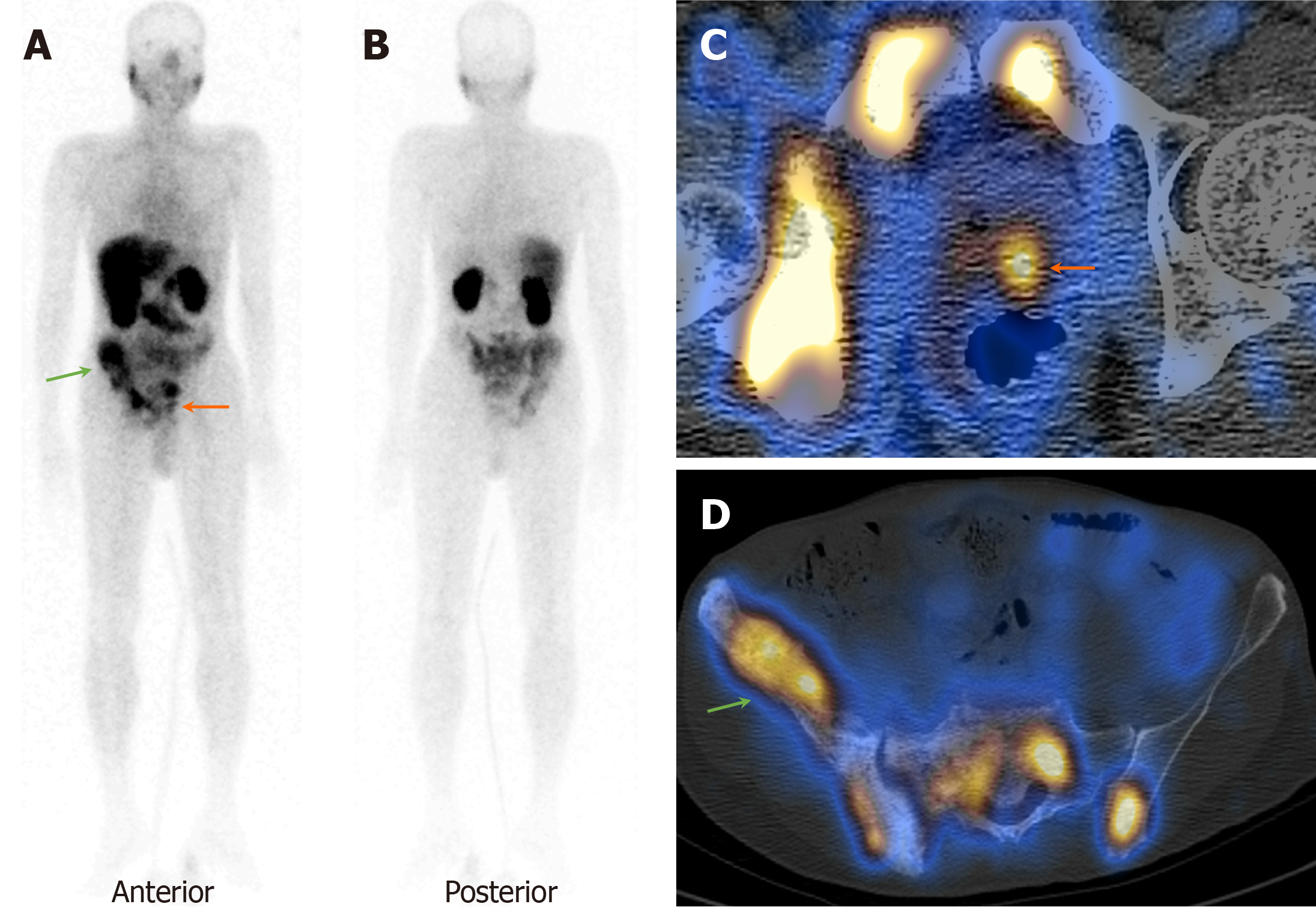
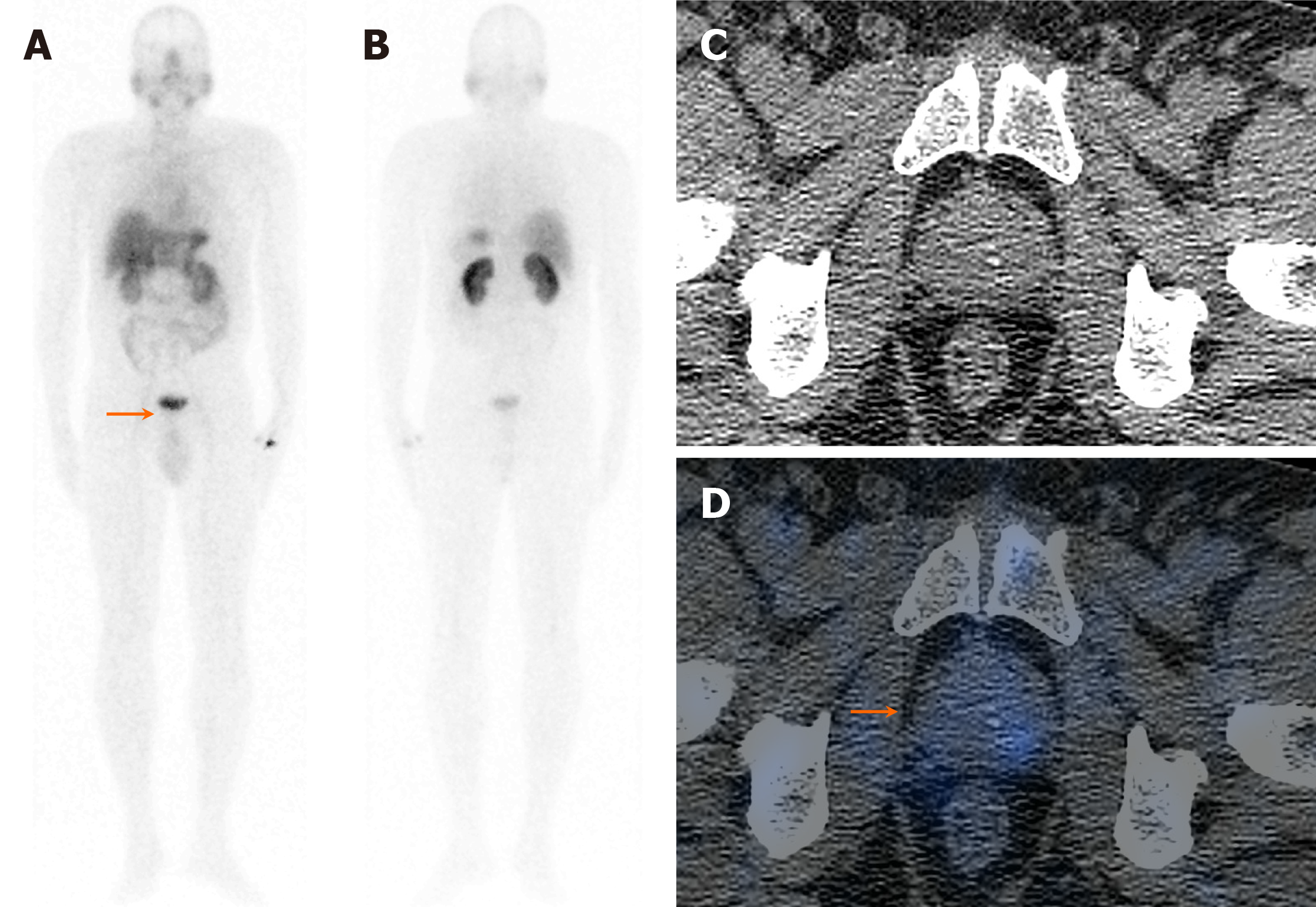
Figure 2 A 64-year-old man presented with difficulty urination and S.
PSA > 100 ng/dL. A and B: 99mTc-PSMA planar images show focal increased tracer uptake in the prostate gland region (orange arrow) and pelvic bones (green arrow); C and D: Axial SPECT-CT fused images show increased focal increased tracer uptake in the left lobe of the prostate gland (orange arrow) and sclerotic pelvic bone lesions (green arrow). Biopsy from the prostate was positive for primary adenocarcinoma (GS-3+3).
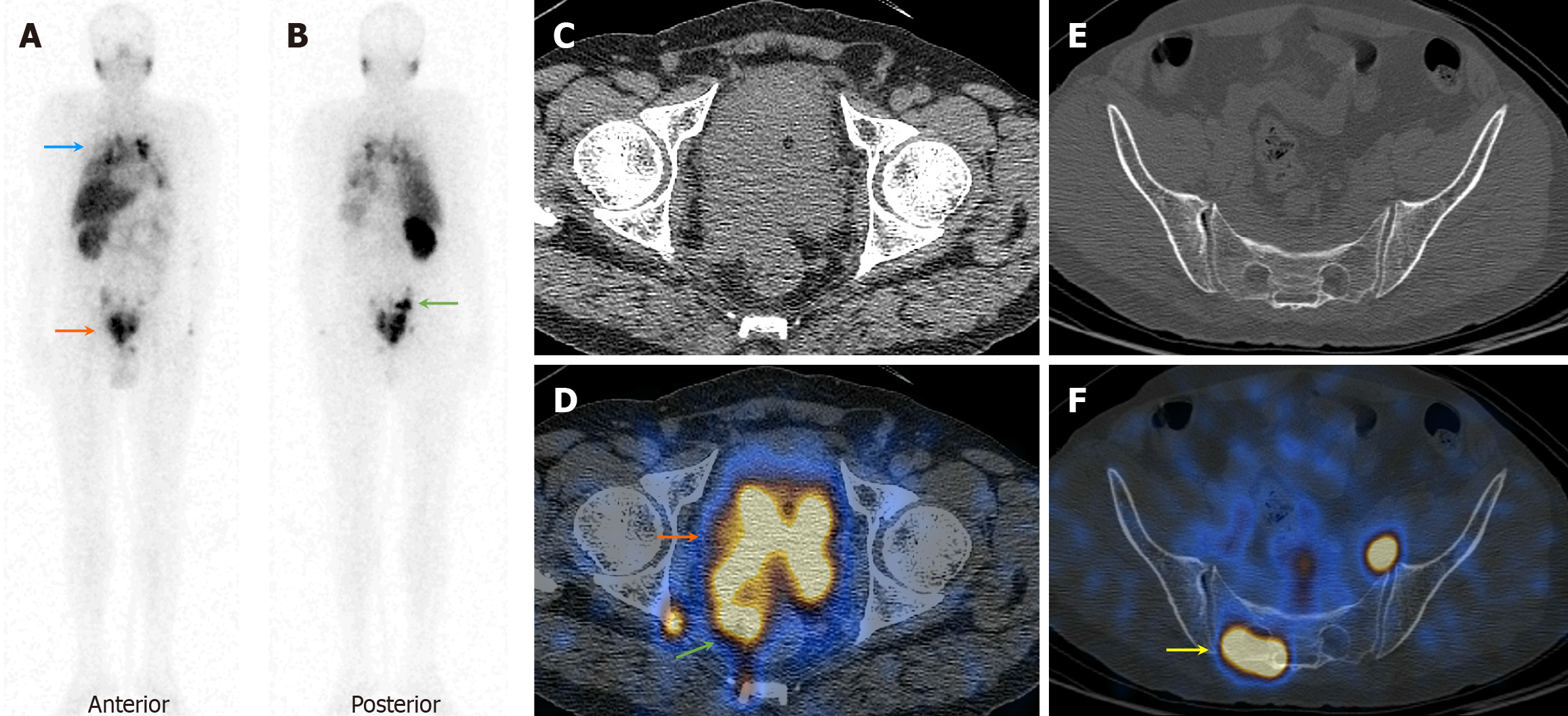
Figure 3 A 65-year-old man presented with difficult and painful micturition, S.
PSA 932 ng/dL, and G-III prostatomegaly on USG. A and B: 99mTc-PSMA planar images show increased heterogenous tracer uptake in the prostate gland region (orange arrow), pelvic nodes (green arrow), and thorax (blue arrow; likely lung and mediastinal nodes); C and D: Axial CT and SPECT-CT fused images show increased tracer uptake in the enlarged prostate gland involving the seminal vesicles, the urinary bladder wall, and the rectum with few pelvic nodes; E and F: Focal increased tracer uptake in the sacrum without corresponding CT lesion (yellow arrow; likely metastatic). Biopsy from the prostate was positive for malignancy.
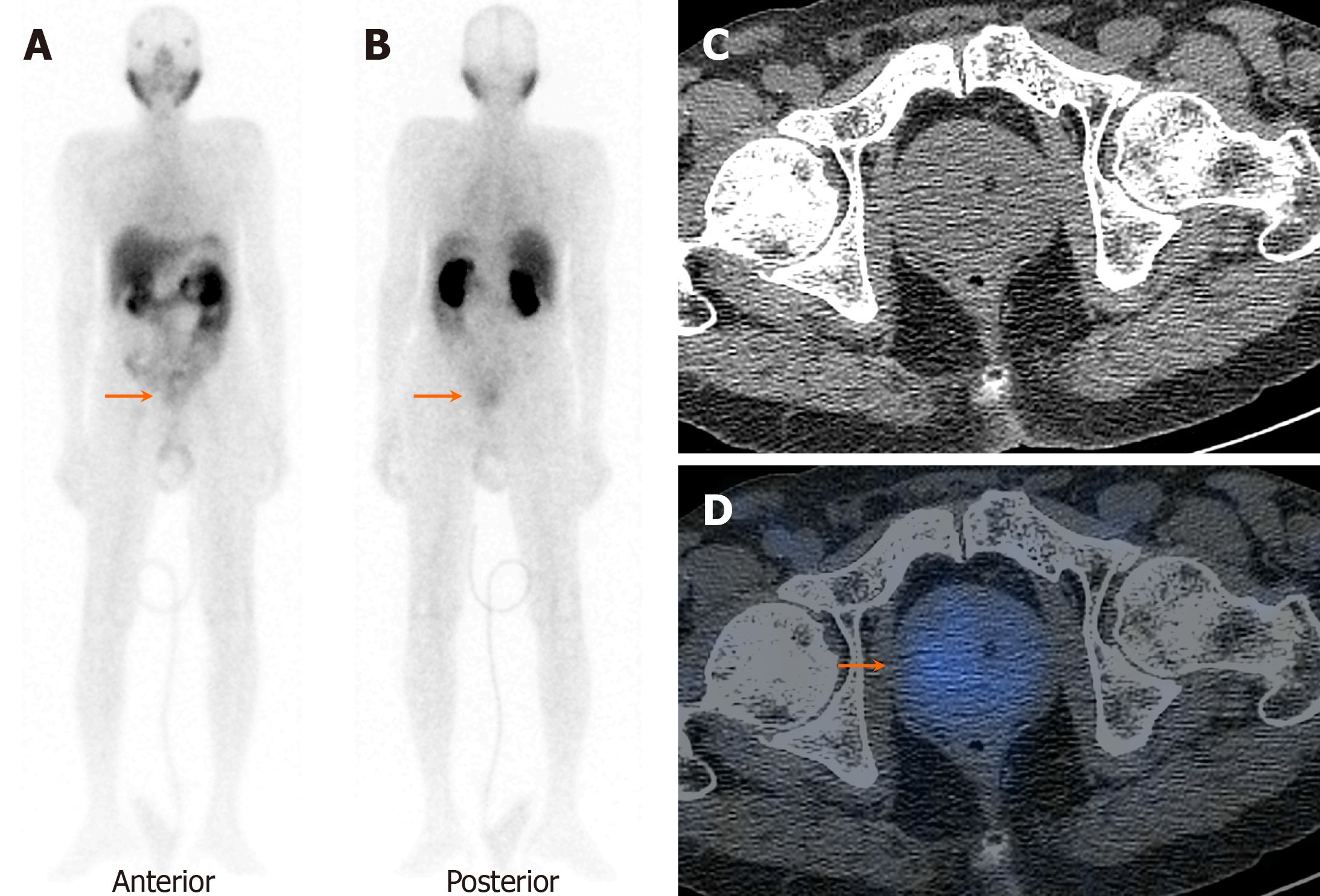
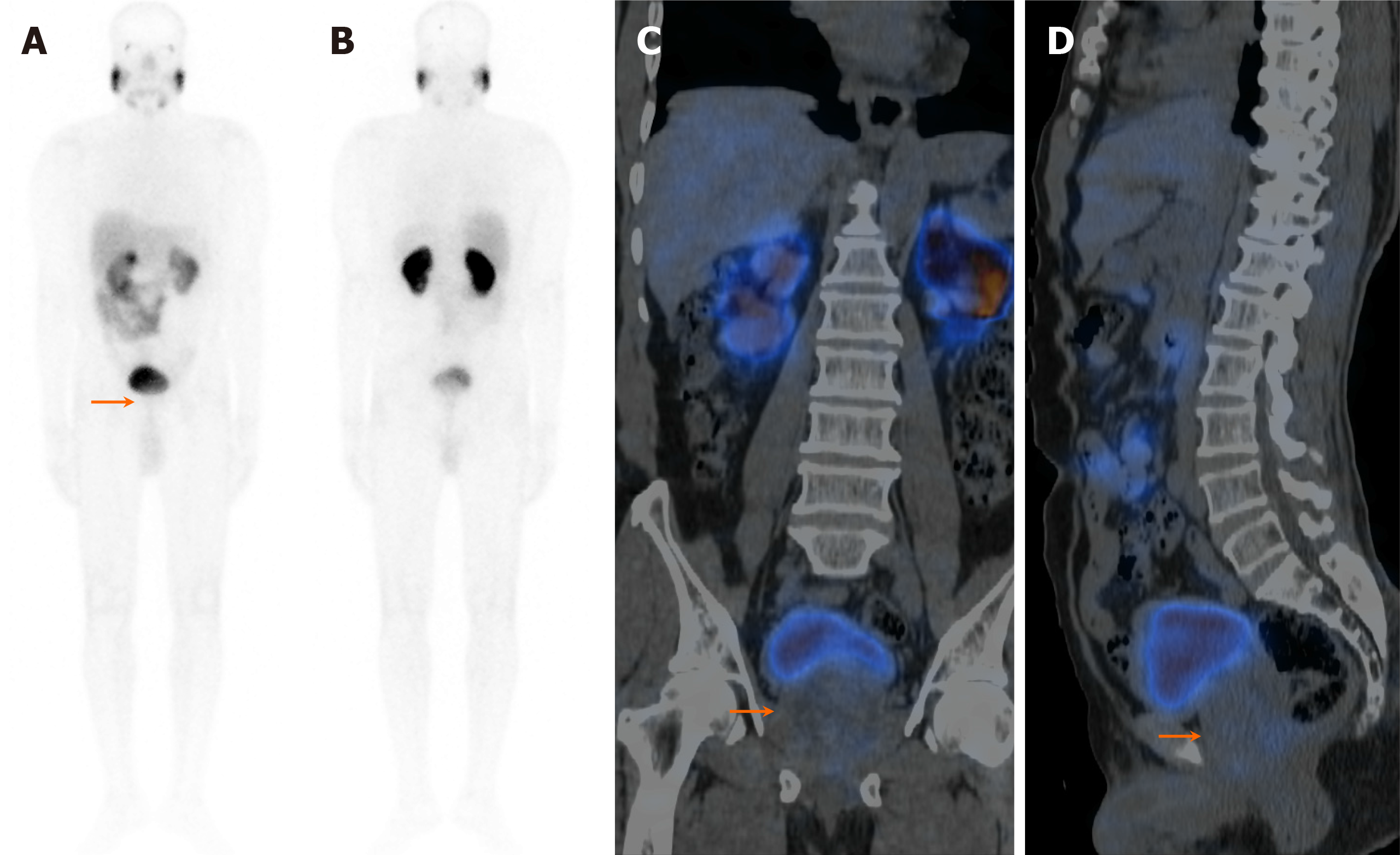
Figure 4 A 75-year-old man presented with hematuria and painful micturition, S.
PSA 48.8 ng/dL, and G-III prostatomegaly on USG. A and B: 99mTc-PSMA planar images show mild diffuse tracer uptake in the prostate gland region (orange arrows), physiological uptake in the salivary glands, liver, spleen, kidneys, and bowel loops, and urinary catheter in situ with no significant bladder activity; C and D: Axial CT and SPECT-CT fused images show prostatomegaly with mild diffuse tracer uptake. Biopsy from the prostate was negative for malignancy with benign hyperplasia.
Figure 5 A 62-year-old man presented with lower urinary tract infections and incomplete voiding, S.
PSA 10.2 ng/dL, and G-IV prosta
Figure 6 A 54-year-old man presented with lower urinary tract infections and difficult micturition, S.
PSA 17.5 ng/dL, and mild prosta
- Citation: Agrawal K, Patro PSS, Singhal T, Satpati D, Nayak P, Mandal S, Kumar N, Padhy BM, Sable M, Meher BR. Diagnostic performance of 99mTc-PSMA SPECT/CT in primary prostate carcinoma. World J Clin Cases 2025; 13(24): 107555
- URL: https://www.wjgnet.com/2307-8960/full/v13/i24/107555.htm
- DOI: https://dx.doi.org/10.12998/wjcc.v13.i24.107555


