Copyright
©The Author(s) 2025.
World J Clin Cases. Aug 26, 2025; 13(24): 107555
Published online Aug 26, 2025. doi: 10.12998/wjcc.v13.i24.107555
Published online Aug 26, 2025. doi: 10.12998/wjcc.v13.i24.107555
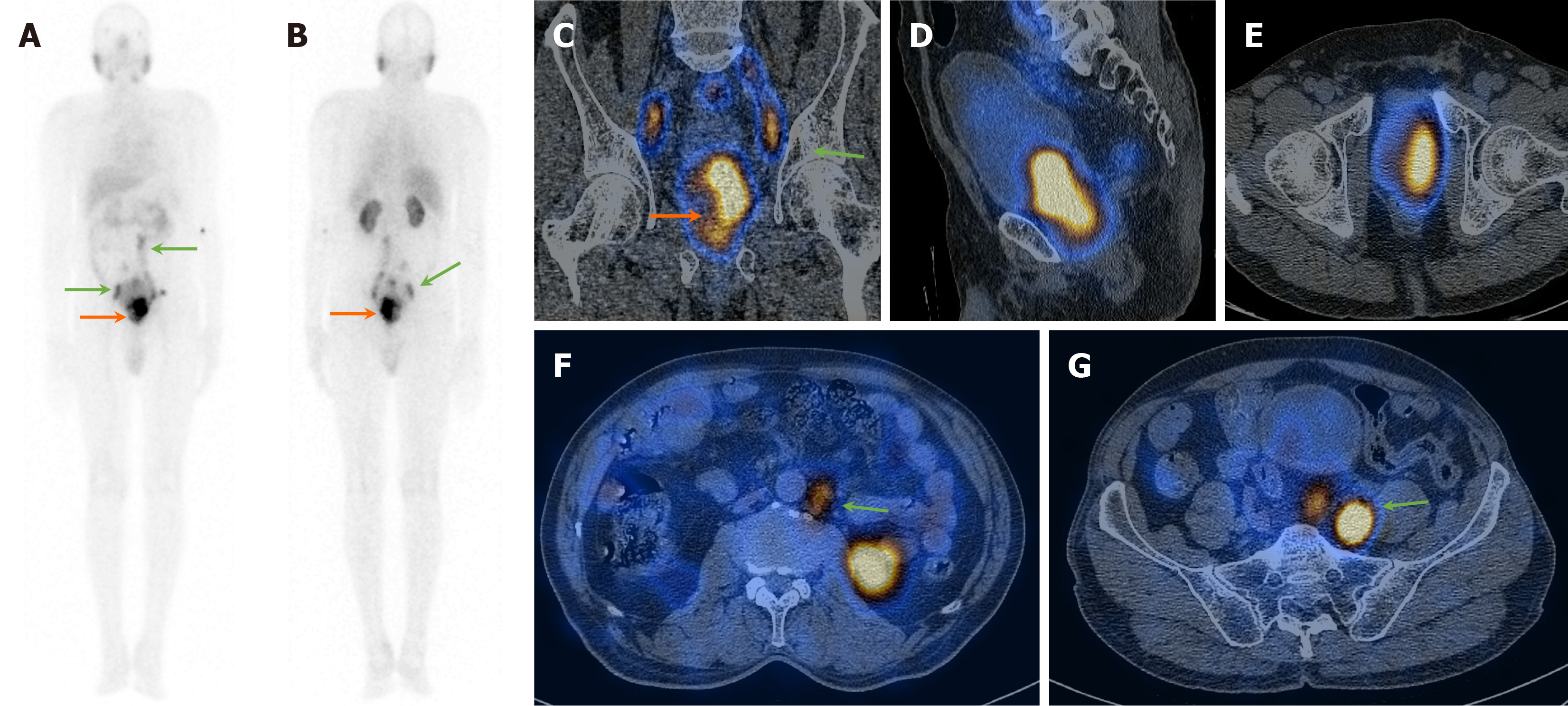
Figure 1 A 59-year-old man presented with hematuria and difficulty urination, S.
PSA > 100 ng/dL, and a suspicious lesion in the prostate on magnetic resonance imaging. A and B: 99mTc-PSMA planar images show increased heterogeneous tracer uptake in the prostate gland region (orange arrows) and likely abdomino-pelvic lymph nodes (green arrows); C-G: Coronal, sagittal, and axial SPECT-CT fused images show increased heterogenous tracer uptake in the enlarged prostate gland (orange arrows) and abdomino-pelvic lymph nodes (green arrows). Biopsy from the prostate was positive for primary adenocarcinoma (GS-5+5).
- Citation: Agrawal K, Patro PSS, Singhal T, Satpati D, Nayak P, Mandal S, Kumar N, Padhy BM, Sable M, Meher BR. Diagnostic performance of 99mTc-PSMA SPECT/CT in primary prostate carcinoma. World J Clin Cases 2025; 13(24): 107555
- URL: https://www.wjgnet.com/2307-8960/full/v13/i24/107555.htm
- DOI: https://dx.doi.org/10.12998/wjcc.v13.i24.107555


