Copyright
©The Author(s) 2025.
World J Clin Cases. Aug 26, 2025; 13(24): 107555
Published online Aug 26, 2025. doi: 10.12998/wjcc.v13.i24.107555
Published online Aug 26, 2025. doi: 10.12998/wjcc.v13.i24.107555
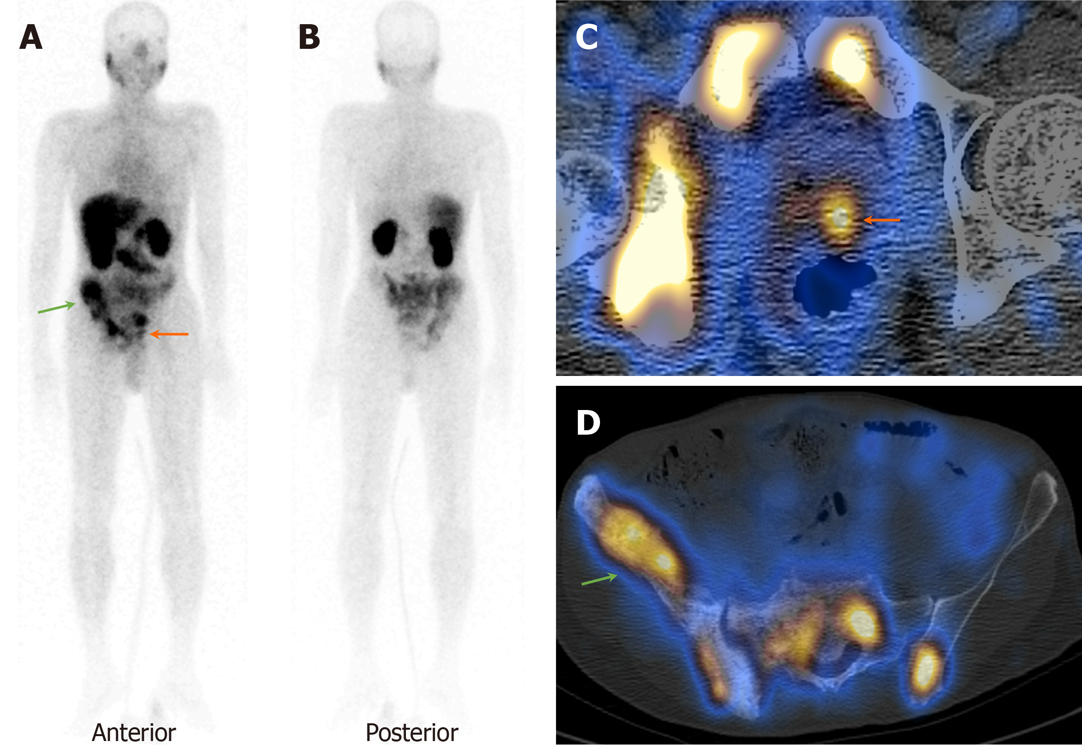
Figure 2 A 64-year-old man presented with difficulty urination and S.
PSA > 100 ng/dL. A and B: 99mTc-PSMA planar images show focal increased tracer uptake in the prostate gland region (orange arrow) and pelvic bones (green arrow); C and D: Axial SPECT-CT fused images show increased focal increased tracer uptake in the left lobe of the prostate gland (orange arrow) and sclerotic pelvic bone lesions (green arrow). Biopsy from the prostate was positive for primary adenocarcinoma (GS-3+3).
- Citation: Agrawal K, Patro PSS, Singhal T, Satpati D, Nayak P, Mandal S, Kumar N, Padhy BM, Sable M, Meher BR. Diagnostic performance of 99mTc-PSMA SPECT/CT in primary prostate carcinoma. World J Clin Cases 2025; 13(24): 107555
- URL: https://www.wjgnet.com/2307-8960/full/v13/i24/107555.htm
- DOI: https://dx.doi.org/10.12998/wjcc.v13.i24.107555


