Copyright
©The Author(s) 2022.
World J Clin Cases. Jan 21, 2022; 10(3): 1093-1098
Published online Jan 21, 2022. doi: 10.12998/wjcc.v10.i3.1093
Published online Jan 21, 2022. doi: 10.12998/wjcc.v10.i3.1093
Figure 1 Preoperative photos of the mass in the superior temporal quadrant of the left eye (case 1).
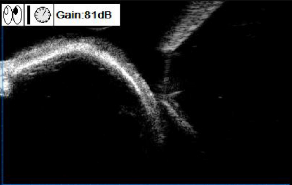
Figure 2 Preoperative ultrasound biomicroscopy image of the mass in the superior temporal quadrant of the left eye (case 1).
A strong oval echo was observed in the superficial sclera under the bulbar conjunctiva, with a clear boundary obscuring the lower echo.
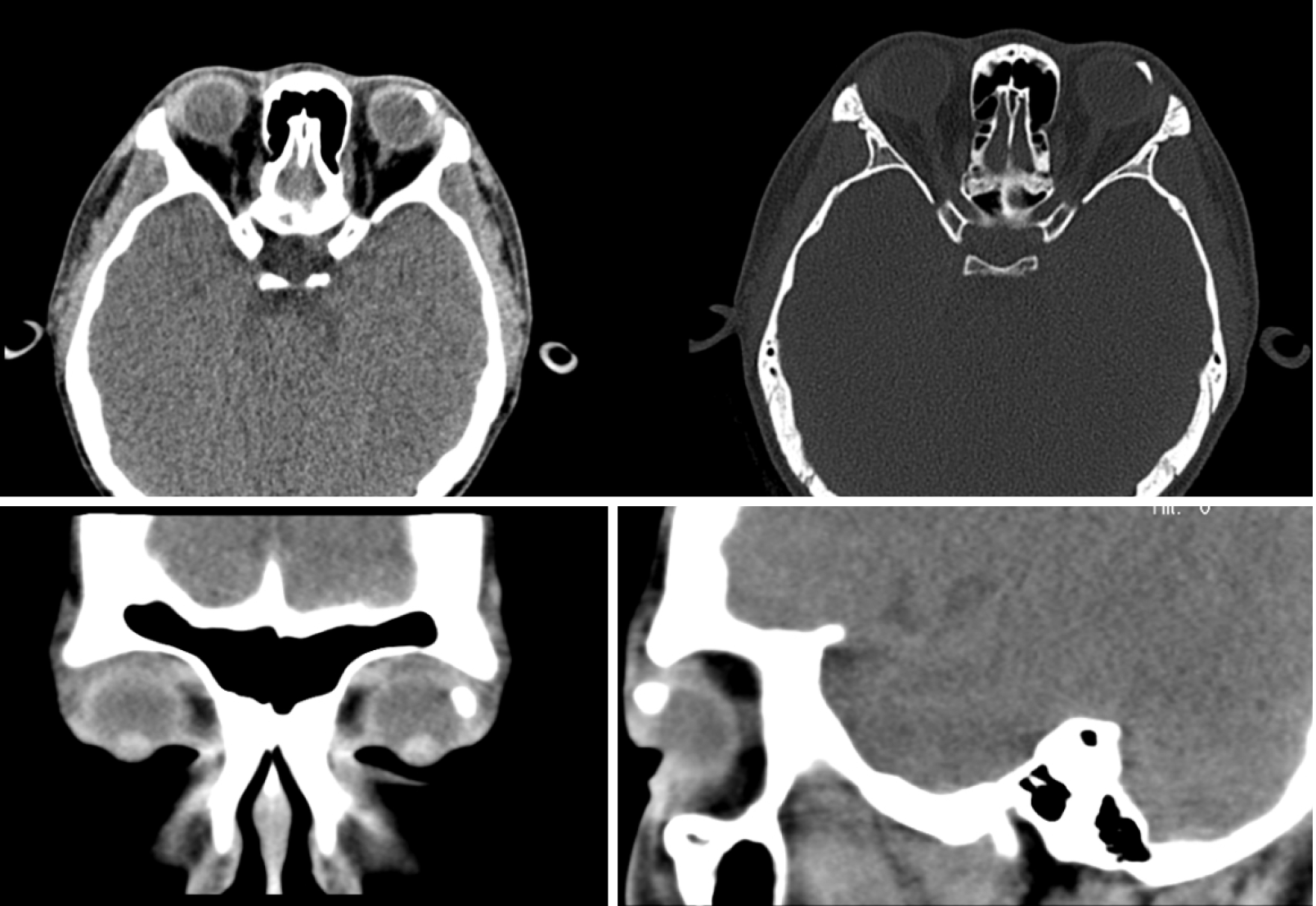
Figure 3 Preoperative computed tomography scan of the mass in the superior temporal quadrant of the left eye (case 1).
Lumps of calcification were apparent in the upper left conjunctiva of the left eye.
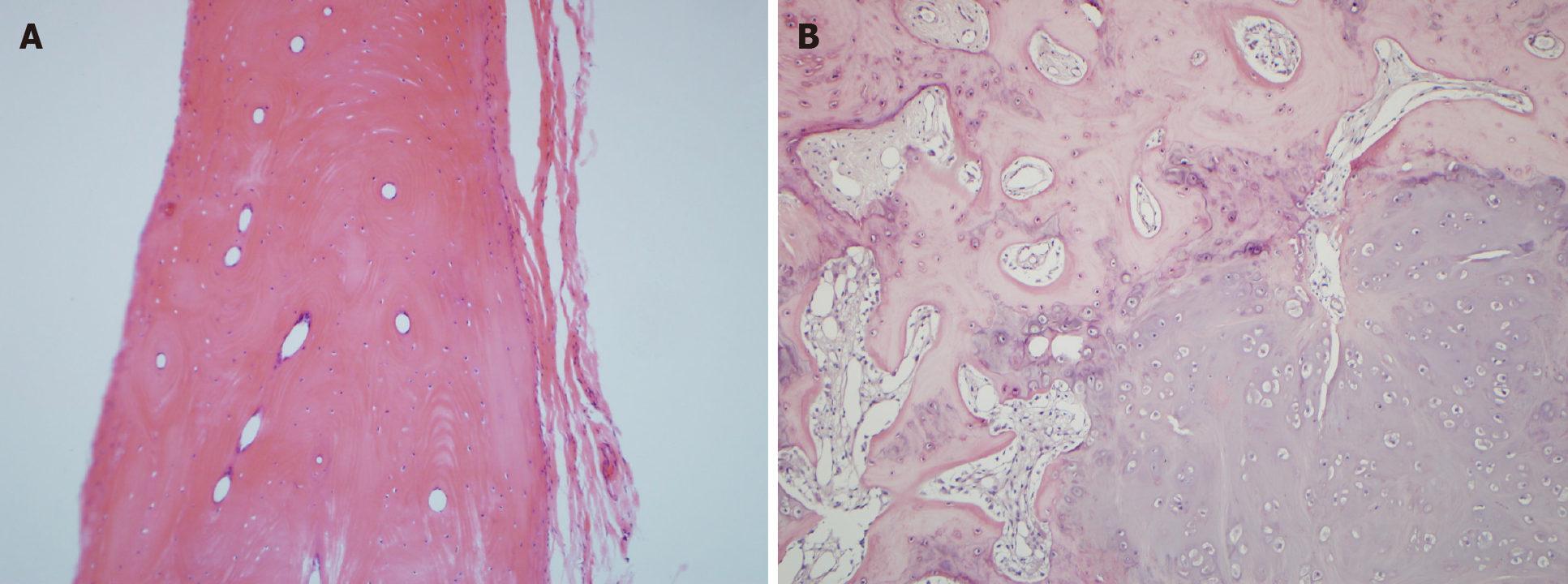
Figure 4 Hematoxylin-eosin staining after resection of the local tumor (magnification, 25 ×).
A: Case 1. The pathology showed features of superficial scleral osteoblastoma: Flat and round tumors that were as hard as bone and surrounded by fibrous connective tissue and fat; B: Case 2. The pathology revealed features of osteoblastoma consisting of bone and cartilage tissue surrounded by numerous collagen fibers.
- Citation: Wang YC, Wang ZZ, You DB, Wang W. Epibulbar osseous choristoma: Two case reports. World J Clin Cases 2022; 10(3): 1093-1098
- URL: https://www.wjgnet.com/2307-8960/full/v10/i3/1093.htm
- DOI: https://dx.doi.org/10.12998/wjcc.v10.i3.1093


