
















Copyright: ©Author(s) 2026.
World J Crit Care Med. Sep 9, 2026; 15(3): 120314
Published online Sep 9, 2026. doi: 10.5492/wjccm.120314
Published online Sep 9, 2026. doi: 10.5492/wjccm.120314
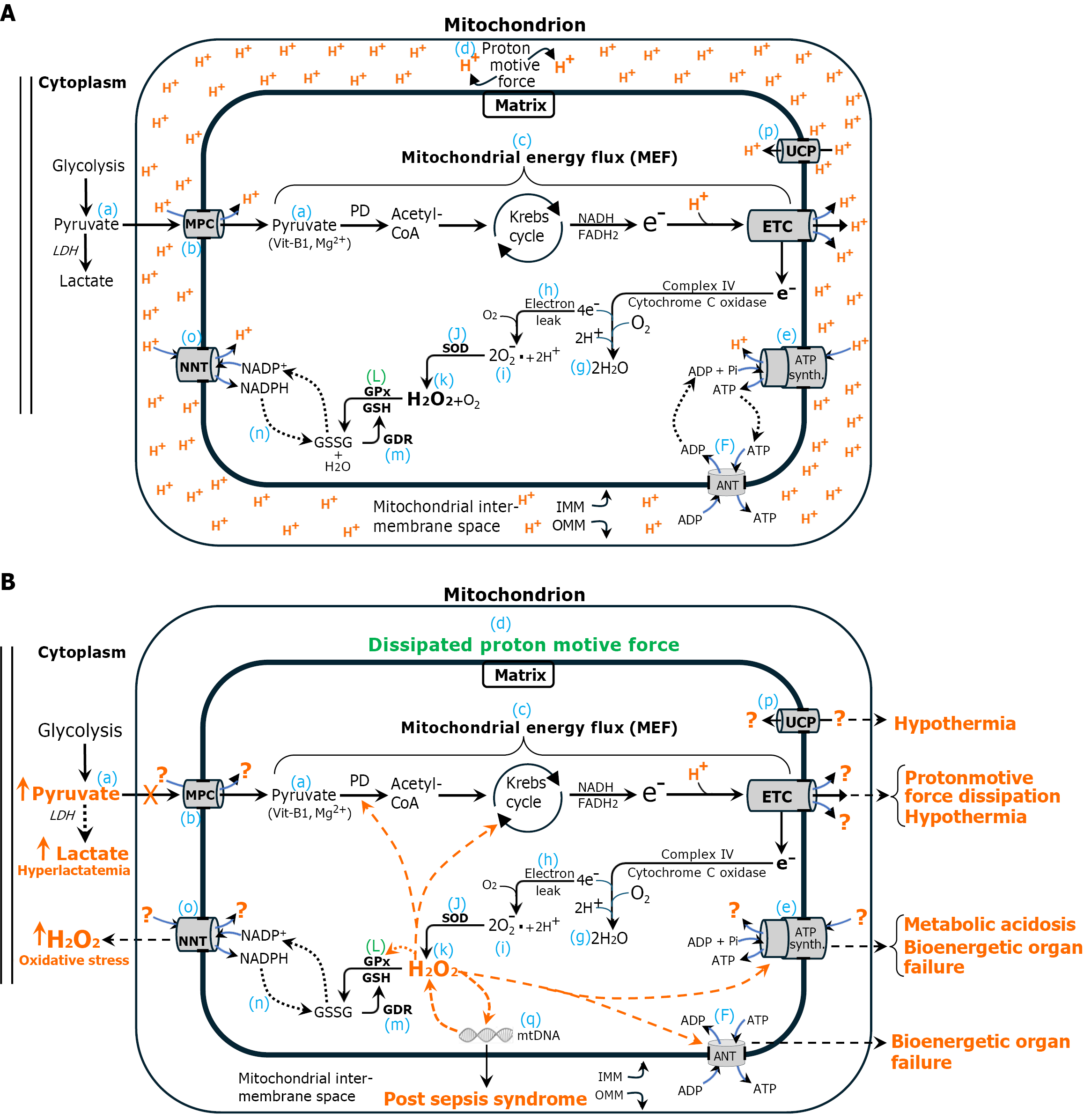
Figure 1 Normal and sepsis bioenergetics.
A: Normal cellular bioenergetics. During normal cellular bioenergetics, cytoplasmic pyruvate (a), the final product of glycolysis, is transported into the mitochondrial matrix via the mitochondrial pyruvate carrier (MPC) (b). There, it undergoes oxidation, beginning with pyruvate dehydrogenase (PD), to generate acetyl CoA, which fuels the Krebs cycle. This process yields NADH and FADH2, which transfer high-energy electrons to the electron transport chain (ETC). The series of sequential enzymatic reactions starting with PD to terminal electron transfer in the ETC complex IV can be visualized as the mitochondrial energy flux (MEF) (c). The ETC utilizes the high-energy electrons to translocate (pump) protons from the mitochondrial matrix into the intermembrane space, establishing the electrochemical proton gradient known as the proton motive force (PMF) (d), whose protons power ATP synthase to synthesize ATP (e). ATP is transported into the cytoplasm by adenine nucleotide translocase (ANT) (F). The MEF is essential for sustaining both ETC function and the PMF. At the terminal step, electrons are transferred to molecular oxygen via ETC complex IV (cytochrome c oxidase), generating water (g). However, up to 4% of electrons prematurely escape into the matrix (electron leak) (h), reducing molecular oxygen to superoxide (i), which is subsequently converted to H2O2 (k) by superoxide dismutase (J). H2O2 is highly toxic and must be immediately degraded. H2O2 is neutralized by glutathione peroxidase (GPx) and glutathione (GSH) (L). The oxidized cofactor GSH disulfide (GSSG) is regenerated to its reduced form (GSH) by GSSG reductase (EC #1.8.1.7) (m) with reducing equivalents provided by NADPH (n). In the process, NADPH is oxidized to NADP+, which is regenerated to NADPH by nicotinamide nucleotide transhydrogenase (NNT) (o) in the inner mitochondrial membrane. NNT is powered by the PMF. Peroxiredoxin and reduced thioredoxin (not shown) are also critical to mitochondrial antioxidant defense (Figure 2). Uncoupling protein (p) dissipates the proton gradient as heat instead of making ATP, reducing energy efficiency to regulate temperature; B: Sepsis bioenergetics: The same as panel A, with the addition of orange dashed arrows to indicate molecular targets inhibited by elevated levels of mitochondrial H2O2, specifically PD and Krebs cycle enzymes. These inhibitory effects compromise mitochondrial energy metabolism and diminish MEF. Consequently, proton translocation into the intermembrane space is impaired, leading to dissipation of the PMF. The loss of PMF disrupts the function of key mitochondrial components, including the MPC, ATP synthase, NNT, and ANT. The terminal metabolic abnormalities associated with each component dysfunction are highlighted in orange text and include elevated lactate levels, increased mitochondrial H2O2, hypothermia, bioenergetic failure, and metabolic acidemia. Each of these parameters serves as a biomarker for the dissipation of the PMF, a critical bioenergetic gradient essential for sustaining life. Their individual associations with increased mortality underscore the fundamental role of PMF integrity in survival. Deficiency of vitamin B1 (thiamine) and magnesium (Mg2+) compromises PD functionality, predisposing to dissipation of the PMF and sepsis. Importantly, correction of these abnormalities in isolation does not improve outcomes in sepsis, as it fails to normalize mitochondrial H2O2 and restore the PMF. Collectively, these findings strongly support the conclusion that dissipation of the PMF represents the proximal and unifying cause of sepsis pathophysiology. Additionally, the figure also illustrates direct inhibition of ATP synthase, ANT, and GPx mediated by elevated levels of H2O2, further exacerbating the previously described metabolic disturbances. H2O2-induced oxidative damage to mitochondrial DNA may play a contributory role in the development of post-sepsis syndrome. Question marks (?) indicate absence of electrons. UCP: Uncoupling protein; MPC: Pyruvate carrier; PD: Pyruvate dehydrogenase; ETC: Electron transport chain; MEF: Mitochondrial energy flux; PMF: Proton motive force; ANT: Adenine nucleotide translocase; GPx: Glutathione peroxidase; GSH: Glutathione; GSSG: Glutathione disulfide; GDR: Glutathione disulfide reductase; OMM: Outer mitochondrial membrane; SOD: Superoxide dismutase; IMM: Inner mitochondrial membrane; NNT: Nucleotide transhydrogenase; Prx: Peroxiredoxin; Trx-r: Thioredoxin; mtDNA: Mitochondrial DNA; H2O2: Hydrogen peroxide.
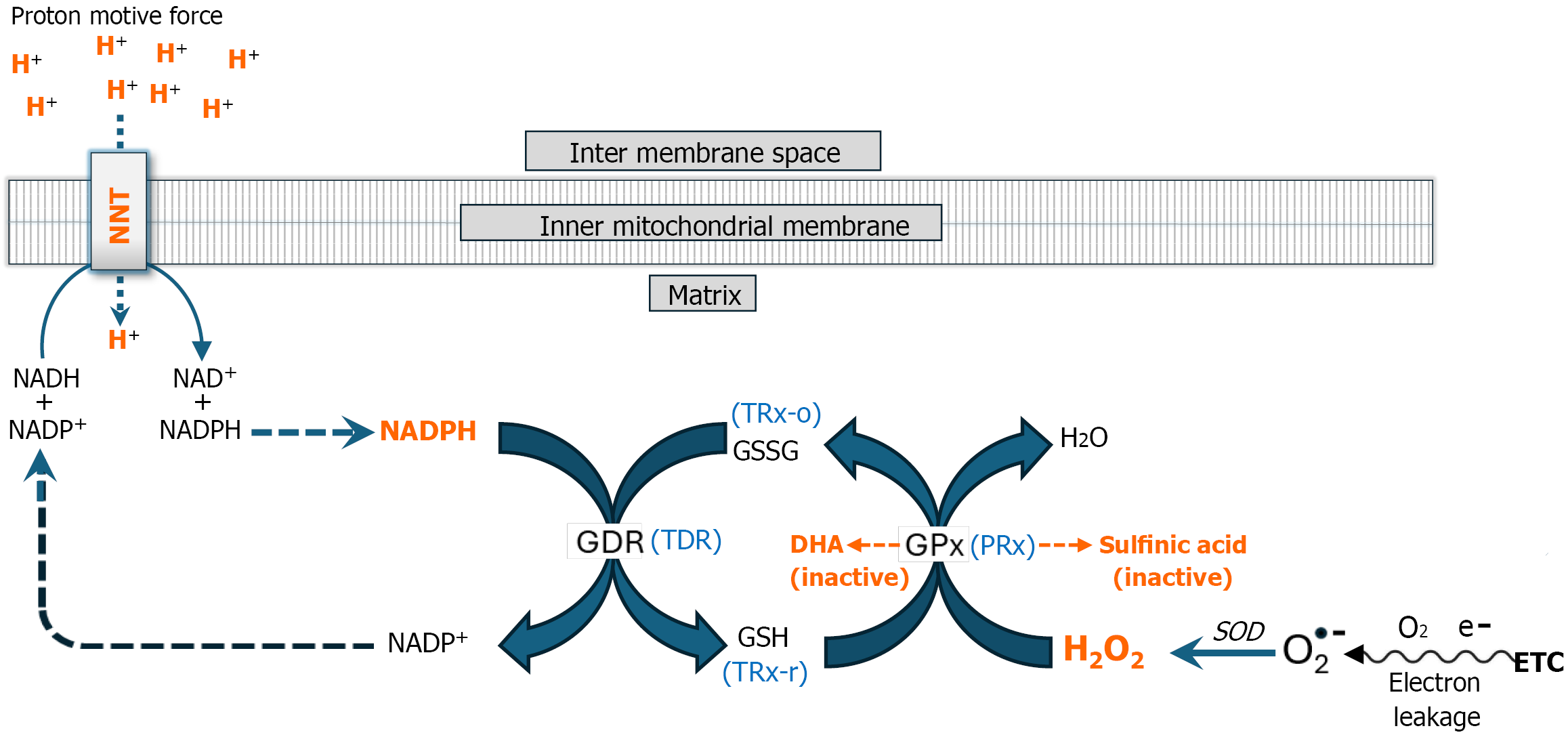
Figure 2 Antioxidant enzyme inhibition: The mitochondrial proton motive force maintains cellular redox homeostasis.
The electron transport chain (ETC) is a major source of hydrogen peroxide (H2O2). Up to 4% of electrons “leak” out prematurely from the ETC into the mitochondrial matrix forming superoxide anion radical (O2•ˉ), which is dismutated to H2O2 by superoxide dismutase. H2O2 is highly toxic to cells and must be disposed of. This detoxification process relies on two key regenerative redox enzyme systems—glutathione peroxidase (GPx) and peroxiredoxin (PRx)—which facilitate the reduction of H2O2 to water using glutathione (GSH) and reduced thioredoxin (TRX-r), respectively. During this reaction, GSH is oxidized to glutathione disulfide (GSSG), while TRX-r is converted to oxidized thioredoxin (TRx-o). To regenerate these reducing agents, glutathione disulfide reductase (EC 1.8.1.7) and thioredoxin disulfide reductase (EC 1.8.1.9) catalyze the reduction of GSSG and TRx-o, respectively, using NADPH as an electron donor. NADPH is replenished by nicotinamide nucleotide transhydrogenase (NNT; EC 7.1.1.1), which facilitates electron transfer from NADH to NADP+. This reaction is driven by the proton motive force (detailed below), linking mitochondrial energy metabolism to redox homeostasis. Thus, mitochondrial enzymatic redox buffering systems critically depend on the proton motive force (PMF) to sustain H2O2 detoxification and maintain cellular redox homeostasis. However, the functionality of this tightly interdependent antioxidant system can be compromised by excessive H2O2 production during the early hypermetabolic stage of sepsis. An acute H2O2 load can deplete GSH and Trx-r, leading to the accumulation of H2O2 followed by hyperoxidation of GPx and PRx, resulting in their conversion to dehydroalanine (DHA) and sulfinic acid derivatives, respectively (orange dashed arrows). These oxidative modifications deactivate both enzymes, which are responsible for eliminating approximately 99% of mitochondrial H2O2. Deactivation of these enzymes results in significantly increased free H2O2 accumulation within the mitochondrion, which disrupts mitochondrial energy metabolism initiating dissipation of the PMF (Figure 1B). Dissipation of the PMF compromises NNT functionality, further reducing mitochondrial capacity for H2O2 detoxification leading to toxic systemic levels of H2O2. NNT: Nucleotide transhydrogenase; GDR: Glutathione disulfide reductase; TDR: Thioredoxin disulfide reductase; GPx: Glutathione peroxidase; PRx: Peroxiredoxin; TRX-r: Reduced thioredoxin; TRx-o: Oxidized thioredoxin; GSH: Glutathione; GSSG: Glutathione disulfide; ETC: Electron transport chain; SOD: Superoxide dismutase; H2O2: Hydrogen peroxide; DHA: Dehydroalanine.
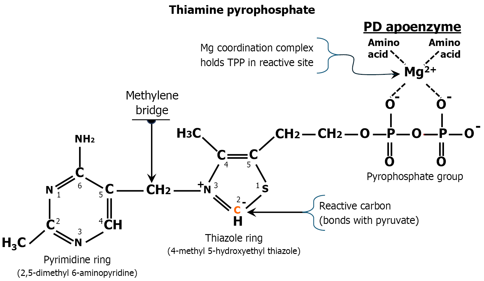
Figure 3 How thiamine or magnesium deficiency predispose to sepsis.
Thiamine (vitamin B1) is transported into cells where it is phosphorylated in the cytoplasm by the addition of two phosphate groups that chelate magnesium (Mg2+) ions. The resulting thiamine pyrophosphate (TPP) is transported into mitochondria where the pyrimidine and thiazole rings provide spatial orientation to localize TPP in the reactive site of the pyruvate dehydrogenase apoenzyme complex (PD apoenzyme), after which the magnesium ions form a coordination complex between peptide side chains of the apoenzyme and the pyrophosphate groups to lock the molecule in place within the reactive site. The precise spatial positioning of thiamine in the reactive site is critical for the subsequent interaction with a negatively charged peptide side chain, which abstracts a proton from the #2 reactive carbon of thiamine’s thiazole ring. The resulting negatively charged reactive carbanion in thiazole’s ring then bonds with pyruvate forming a covalent stabilized transition-state TPP-acetyl complex. This fixation within the active site allows pyruvate to undergo enzymatic oxidative decarboxylation by pyruvate dehydrogenase with the release of one molecule of acetyl-CoA, one molecule of NADH and carbon dioxide gas. The resulting acetyl-CoA then enters the Krebs cycle, where it generates three additional NADH and one FADH2 per cycle. Thiamine is essential for this process to occur, as its absence prevents pyruvate dehydrogenase from capturing pyruvate. Without thiamine, acetyl-CoA synthesis is impaired, disrupting the Krebs cycle and inhibiting the production of NADH and FADH2. This de-energizes the mitochondrial energy flux followed by dissipation of the proton motive force, which compromises the mitochondrial pyruvate carrier and ATP synthase. These disruptions contribute to hyperlactatemia and bioenergetic failure, respectively. Similarly, magnesium deficiency inhibits the binding of thiamine pyrophosphate at the pyruvate dehydrogenase apoenzyme reactive site, resulting in a functional thiamine deficiency despite sufficient thiamine availability. Consequently, thiamine or magnesium deficiency increases susceptibility to sepsis due to dissipation of the proton motive force, as previously described (Figure 1B). The critical role of thiamine in this metabolic process cannot be bypassed. TPP: Thiamine pyrophosphate; PD: Pyruvate dehydrogenase.
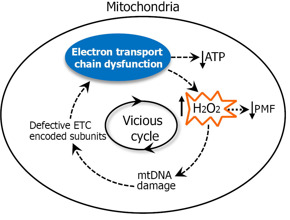
Figure 4 Mitochondrial DNA oxidative damage: A diagrammatic representation illustrating the mutagenic impact of electron transport chain-derived hydrogen peroxide on mitochondrial DNA.
Mitochondrial DNA (mtDNA) is highly susceptible to oxidative damage. Exposure to hydrogen peroxide (H2O2) during sepsis leads to mtDNA oxidative damage. This damage gives rise to mtDNA mutations (mitochondrial heteroplasmy), resulting in transcriptional miscoding of electron transport chain (ETC) complexes and the subsequent synthesis of defective and mutated ETC protein subunits. These dysfunctional proteins disrupt electron flow, causing an elevated rate of electron leakage. The prematurely ‘leaked’ electrons interact with nearby molecular oxygen within the mitochondrial matrix, generating superoxide, which is subsequently converted to H2O2 through the action of superoxide dismutase. The H2O2 leads to additional mtDNA damage resulting in an iterative cycle of oxidative mtDNA damage, defective ETC protein complexes and progressively greater levels of H2O2. This disruption initiates a self-amplifying (vicious) cycle of increasing H2O2 production and ETC dysfunction while compromising oxidative phosphorylation and ATP synthesis. The elevated H2O2 levels inhibit the mitochondrial energy flux and dissipate the proton motive force while simultaneously contributing to immuno
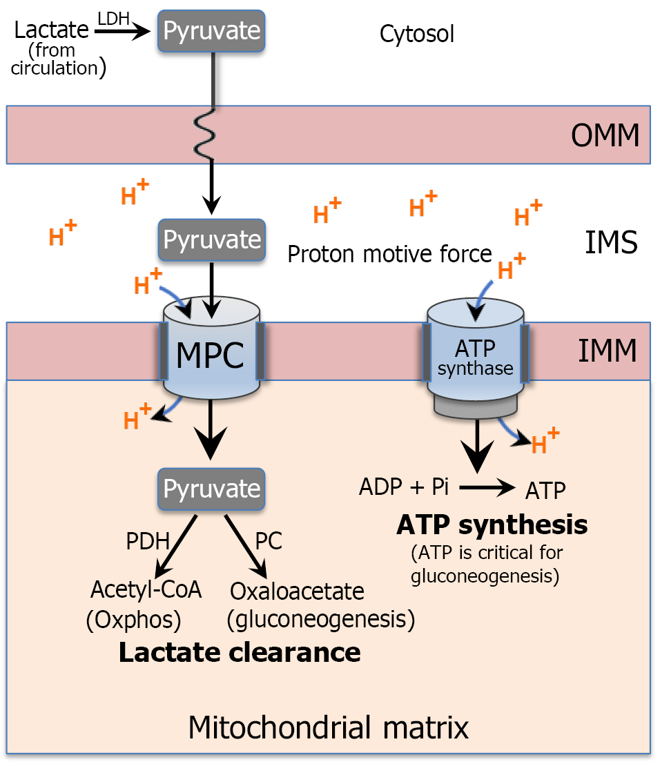
Figure 5 Hepatic lactate clearance depends on the proton motive force: Hepatic clearance of serum lactate requires its reduction to pyruvate within hepatocytes, followed by either conversion of pyruvate to glucose through gluconeogenesis (the Cori cycle) or oxidation of pyruvate after its transport into mitochondria.
Gluconeogenesis depends on an intact proton motive force (PMF) because pyruvate must be transported into the mitochondrial matrix by the mitochondrial pyruvate carrier (mitochondrial phase of gluconeogenesis), a process driven directly by the PMF. Dissipation of the PMF impairs this transport step, blocking both pyruvate oxidation and the mitochondrial phase of gluconeogenesis. PMF loss also reduces ATP synthesis by ATP synthase, limiting the ATP required for several energy dependent reactions in the gluconeogenesis pathway. Each glucose molecule produced requires 2 lactate molecules and consumes 6 ATP equivalents, linking gluconeogenesis tightly to mitochondrial bioenergetics. Therefore, hepatic lactate clearance is fundamentally dependent on the integrity of the proton motive force, and its dissipation can lead to impaired lactate metabolism (clearance) and subsequent hyperlactatemia. LDH: Lactate dehydrogenase; OMM: Outer mitochondrial membrane; IMS: Mitochondrial intermembrane space; MPC: Pyruvate carrier; IMM: Inner mitochondrial membrane; PD: Pyruvate dehydrogenase; PC: Pyruvate carboxylase.
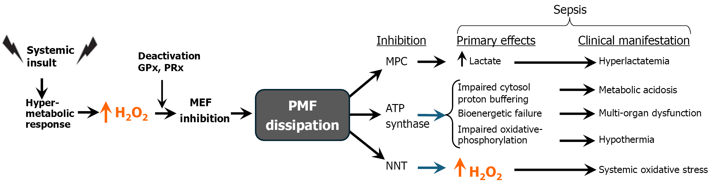
Figure 6 Overview of Sepsis Pathogenesis.
A systemic insult triggers a hypermetabolic response that markedly elevates mitochondrial hydrogen peroxide (H2O2) production. When H2O2 concentrations exceed the buffering capacity of key intracellular reducing agents—namely glutathione and reduced thioredoxin—accumulation of free H2O2 ensues. This excess H2O2 inactivates essential antioxidant enzymes, including glutathione peroxidase and peroxiredoxin, thereby increasing H2O2 within the mitochondria. The resulting H2O2-induced oxidative burden impairs mitochondrial metabolism by inhibiting pyruvate dehydrogenase, key enzymes of the Krebs cycle, and complex II of the electron transport chain. These disruptions compromise mitochondrial energy metabolism (mitochondrial energy flux), impeding the synthesis of critical electron carriers NADH and FADH2. In the absence of these high-energy intermediates, the electrochemical proton gradient cannot be sustained, leading to dissipation of the proton motive force (PMF). Loss of PMF directly impairs the function of inner mitochondrial membrane proteins—including the mitochondrial pyruvate carrier, ATP synthase, and nicotinamide nucleotide transhydrogenase—all of which are powered by PMF. Dysfunction of these components contributes to the hallmark clinical manifestations of sepsis. The elevated mortality associated with these metabolic derangements is a reflection of a dissipated PMF and underscores the essential role of the PMF in sustaining cellular viability. MEF: Mitochondrial energy flux; PMF: Proton motive force; MPC: Pyruvate carrier; NNT: Nucleotide transhydrogenase; H2O2: Hydrogen peroxide.
- Citation: Pravda J. Dissipation of the mitochondrial proton motive force drives sepsis pathogenesis and explains hyperlactatemia’s predictive value in sepsis mortality. World J Crit Care Med 2026; 15(3): 120314
- URL: https://www.wjgnet.com/2220-3141/full/v15/i3/120314.htm
- DOI: https://dx.doi.org/10.5492/wjccm.120314