
















Copyright: ©Author(s) 2026.
World J Crit Care Med. Sep 9, 2026; 15(3): 120314
Published online Sep 9, 2026. doi: 10.5492/wjccm.120314
Published online Sep 9, 2026. doi: 10.5492/wjccm.120314
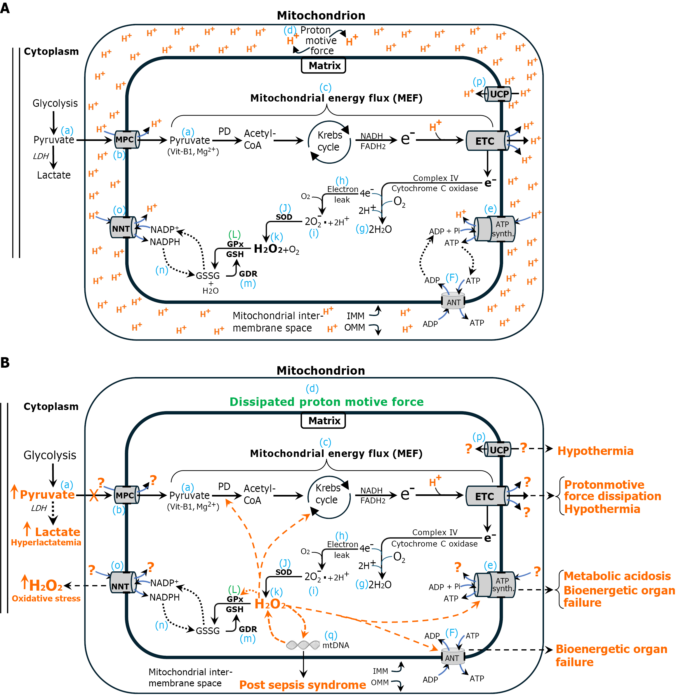
Figure 1 Normal and sepsis bioenergetics.
A: Normal cellular bioenergetics. During normal cellular bioenergetics, cytoplasmic pyruvate (a), the final product of glycolysis, is transported into the mitochondrial matrix via the mitochondrial pyruvate carrier (MPC) (b). There, it undergoes oxidation, beginning with pyruvate dehydrogenase (PD), to generate acetyl CoA, which fuels the Krebs cycle. This process yields NADH and FADH2, which transfer high-energy electrons to the electron transport chain (ETC). The series of sequential enzymatic reactions starting with PD to terminal electron transfer in the ETC complex IV can be visualized as the mitochondrial energy flux (MEF) (c). The ETC utilizes the high-energy electrons to translocate (pump) protons from the mitochondrial matrix into the intermembrane space, establishing the electrochemical proton gradient known as the proton motive force (PMF) (d), whose protons power ATP synthase to synthesize ATP (e). ATP is transported into the cytoplasm by adenine nucleotide translocase (ANT) (F). The MEF is essential for sustaining both ETC function and the PMF. At the terminal step, electrons are transferred to molecular oxygen via ETC complex IV (cytochrome c oxidase), generating water (g). However, up to 4% of electrons prematurely escape into the matrix (electron leak) (h), reducing molecular oxygen to superoxide (i), which is subsequently converted to H2O2 (k) by superoxide dismutase (J). H2O2 is highly toxic and must be immediately degraded. H2O2 is neutralized by glutathione peroxidase (GPx) and glutathione (GSH) (L). The oxidized cofactor GSH disulfide (GSSG) is regenerated to its reduced form (GSH) by GSSG reductase (EC #1.8.1.7) (m) with reducing equivalents provided by NADPH (n). In the process, NADPH is oxidized to NADP+, which is regenerated to NADPH by nicotinamide nucleotide transhydrogenase (NNT) (o) in the inner mitochondrial membrane. NNT is powered by the PMF. Peroxiredoxin and reduced thioredoxin (not shown) are also critical to mitochondrial antioxidant defense (Figure 2). Uncoupling protein (p) dissipates the proton gradient as heat instead of making ATP, reducing energy efficiency to regulate temperature; B: Sepsis bioenergetics: The same as panel A, with the addition of orange dashed arrows to indicate molecular targets inhibited by elevated levels of mitochondrial H2O2, specifically PD and Krebs cycle enzymes. These inhibitory effects compromise mitochondrial energy metabolism and diminish MEF. Consequently, proton translocation into the intermembrane space is impaired, leading to dissipation of the PMF. The loss of PMF disrupts the function of key mitochondrial components, including the MPC, ATP synthase, NNT, and ANT. The terminal metabolic abnormalities associated with each component dysfunction are highlighted in orange text and include elevated lactate levels, increased mitochondrial H2O2, hypothermia, bioenergetic failure, and metabolic acidemia. Each of these parameters serves as a biomarker for the dissipation of the PMF, a critical bioenergetic gradient essential for sustaining life. Their individual associations with increased mortality underscore the fundamental role of PMF integrity in survival. Deficiency of vitamin B1 (thiamine) and magnesium (Mg2+) compromises PD functionality, predisposing to dissipation of the PMF and sepsis. Importantly, correction of these abnormalities in isolation does not improve outcomes in sepsis, as it fails to normalize mitochondrial H2O2 and restore the PMF. Collectively, these findings strongly support the conclusion that dissipation of the PMF represents the proximal and unifying cause of sepsis pathophysiology. Additionally, the figure also illustrates direct inhibition of ATP synthase, ANT, and GPx mediated by elevated levels of H2O2, further exacerbating the previously described metabolic disturbances. H2O2-induced oxidative damage to mitochondrial DNA may play a contributory role in the development of post-sepsis syndrome. Question marks (?) indicate absence of electrons. UCP: Uncoupling protein; MPC: Pyruvate carrier; PD: Pyruvate dehydrogenase; ETC: Electron transport chain; MEF: Mitochondrial energy flux; PMF: Proton motive force; ANT: Adenine nucleotide translocase; GPx: Glutathione peroxidase; GSH: Glutathione; GSSG: Glutathione disulfide; GDR: Glutathione disulfide reductase; OMM: Outer mitochondrial membrane; SOD: Superoxide dismutase; IMM: Inner mitochondrial membrane; NNT: Nucleotide transhydrogenase; Prx: Peroxiredoxin; Trx-r: Thioredoxin; mtDNA: Mitochondrial DNA; H2O2: Hydrogen peroxide.
- Citation: Pravda J. Dissipation of the mitochondrial proton motive force drives sepsis pathogenesis and explains hyperlactatemia’s predictive value in sepsis mortality. World J Crit Care Med 2026; 15(3): 120314
- URL: https://www.wjgnet.com/2220-3141/full/v15/i3/120314.htm
- DOI: https://dx.doi.org/10.5492/wjccm.120314