
















Copyright: ©Author(s) 2026.
World J Crit Care Med. Sep 9, 2026; 15(3): 120314
Published online Sep 9, 2026. doi: 10.5492/wjccm.120314
Published online Sep 9, 2026. doi: 10.5492/wjccm.120314
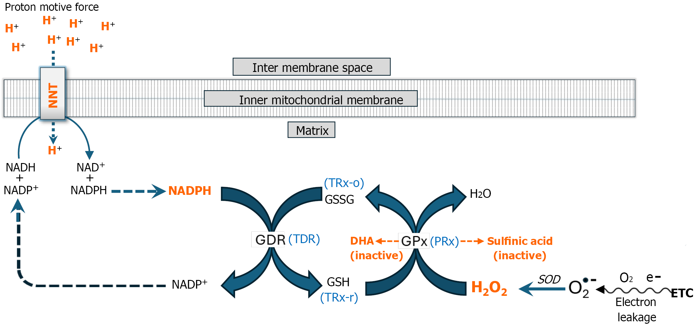
Figure 2 Antioxidant enzyme inhibition: The mitochondrial proton motive force maintains cellular redox homeostasis.
The electron transport chain (ETC) is a major source of hydrogen peroxide (H2O2). Up to 4% of electrons “leak” out prematurely from the ETC into the mitochondrial matrix forming superoxide anion radical (O2•ˉ), which is dismutated to H2O2 by superoxide dismutase. H2O2 is highly toxic to cells and must be disposed of. This detoxification process relies on two key regenerative redox enzyme systems—glutathione peroxidase (GPx) and peroxiredoxin (PRx)—which facilitate the reduction of H2O2 to water using glutathione (GSH) and reduced thioredoxin (TRX-r), respectively. During this reaction, GSH is oxidized to glutathione disulfide (GSSG), while TRX-r is converted to oxidized thioredoxin (TRx-o). To regenerate these reducing agents, glutathione disulfide reductase (EC 1.8.1.7) and thioredoxin disulfide reductase (EC 1.8.1.9) catalyze the reduction of GSSG and TRx-o, respectively, using NADPH as an electron donor. NADPH is replenished by nicotinamide nucleotide transhydrogenase (NNT; EC 7.1.1.1), which facilitates electron transfer from NADH to NADP+. This reaction is driven by the proton motive force (detailed below), linking mitochondrial energy metabolism to redox homeostasis. Thus, mitochondrial enzymatic redox buffering systems critically depend on the proton motive force (PMF) to sustain H2O2 detoxification and maintain cellular redox homeostasis. However, the functionality of this tightly interdependent antioxidant system can be compromised by excessive H2O2 production during the early hypermetabolic stage of sepsis. An acute H2O2 load can deplete GSH and Trx-r, leading to the accumulation of H2O2 followed by hyperoxidation of GPx and PRx, resulting in their conversion to dehydroalanine (DHA) and sulfinic acid derivatives, respectively (orange dashed arrows). These oxidative modifications deactivate both enzymes, which are responsible for eliminating approximately 99% of mitochondrial H2O2. Deactivation of these enzymes results in significantly increased free H2O2 accumulation within the mitochondrion, which disrupts mitochondrial energy metabolism initiating dissipation of the PMF (Figure 1B). Dissipation of the PMF compromises NNT functionality, further reducing mitochondrial capacity for H2O2 detoxification leading to toxic systemic levels of H2O2. NNT: Nucleotide transhydrogenase; GDR: Glutathione disulfide reductase; TDR: Thioredoxin disulfide reductase; GPx: Glutathione peroxidase; PRx: Peroxiredoxin; TRX-r: Reduced thioredoxin; TRx-o: Oxidized thioredoxin; GSH: Glutathione; GSSG: Glutathione disulfide; ETC: Electron transport chain; SOD: Superoxide dismutase; H2O2: Hydrogen peroxide; DHA: Dehydroalanine.
- Citation: Pravda J. Dissipation of the mitochondrial proton motive force drives sepsis pathogenesis and explains hyperlactatemia’s predictive value in sepsis mortality. World J Crit Care Med 2026; 15(3): 120314
- URL: https://www.wjgnet.com/2220-3141/full/v15/i3/120314.htm
- DOI: https://dx.doi.org/10.5492/wjccm.120314