Copyright
©The Author(s) 2025.
World J Orthop. Aug 18, 2025; 16(8): 109904
Published online Aug 18, 2025. doi: 10.5312/wjo.v16.i8.109904
Published online Aug 18, 2025. doi: 10.5312/wjo.v16.i8.109904
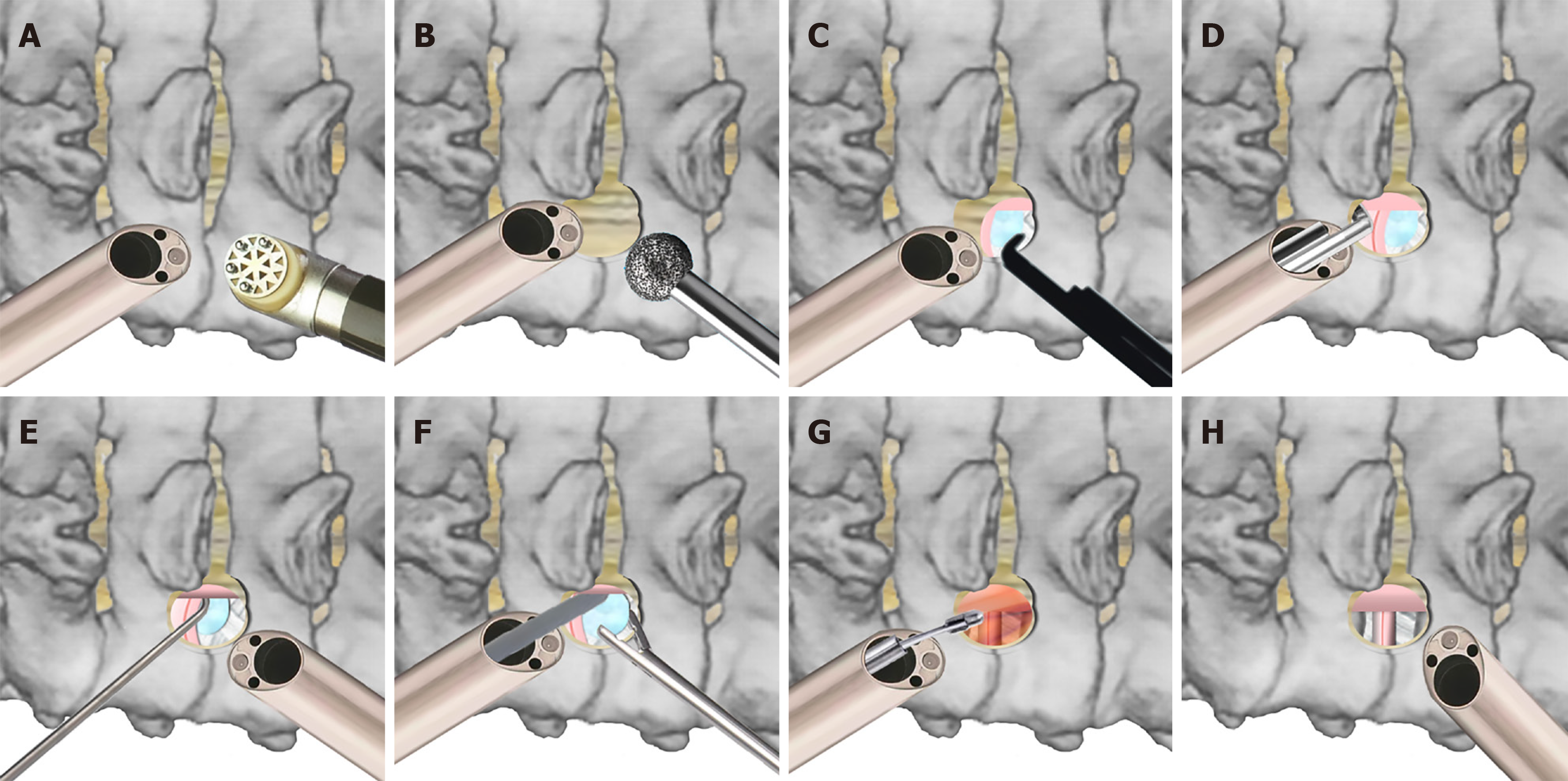
Figure 1 Schematic diagram of hybrid technique technology operation.
A: Extraspinal soft tissue exfoliation and hemostasis by high-energy radiofrequency probe [unilateral biportal endoscopy (UBE) mode]; B: Laminotomy by endoscopic drill (UBE mode); C: Preliminary ligamentum flavum resection by regular punch (UBE mode); D: Precise resection of ligamentum flavum endoscopic punch [uniportal endoscopic surgery (UES) mode]; E: Exchange portals and explore intraspinal tissue by nerve hook(Interchanged mode); F: Remove herniated nucleus pulposus by regular pituitary forceps and tract nerve root by stripper (hybrid technique mode); G: Intraspinal hemostasis using low-energy radiofrequency probe (UES mode); H: Examination before closure under enlarged surgical field (interchanged mode).
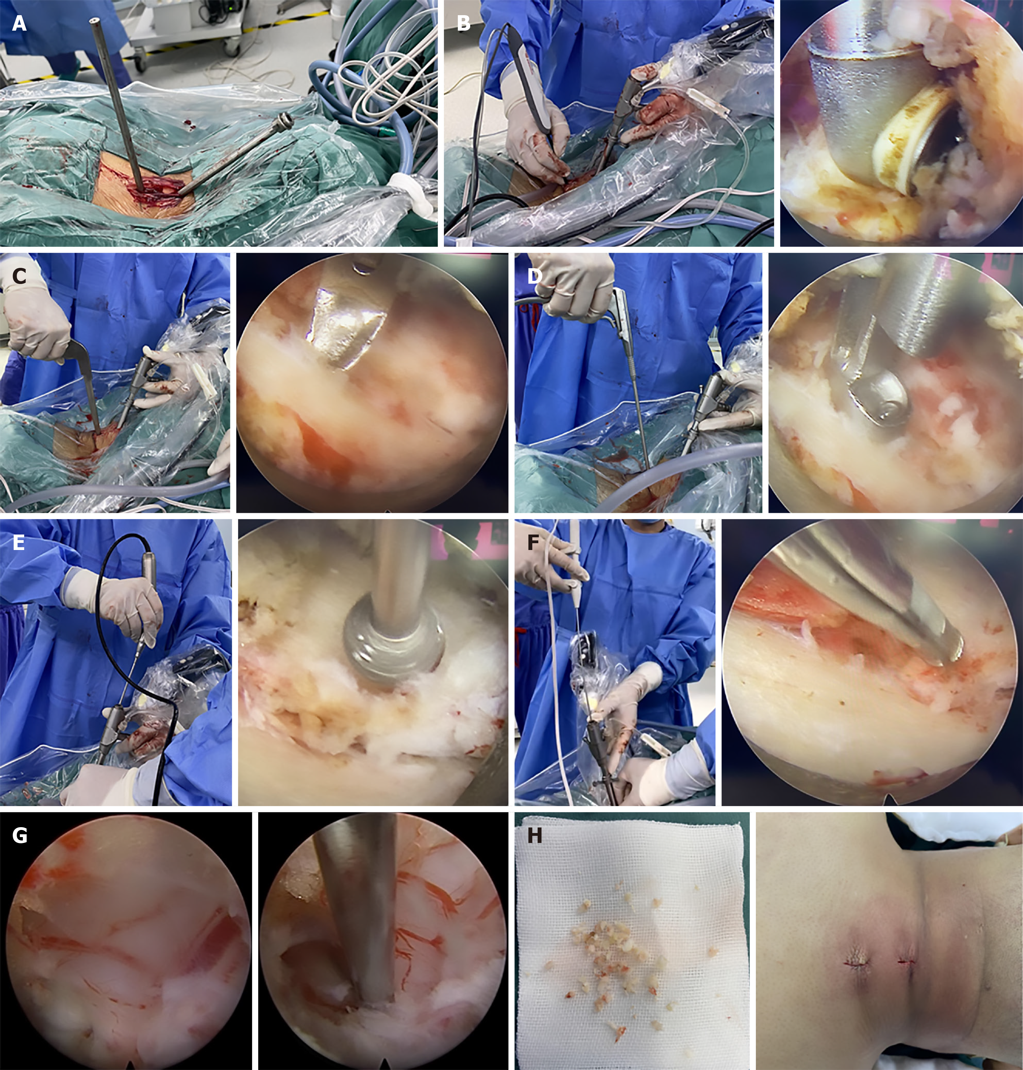
Figure 2 Main procedures in hybrid technique group.
A: Set-up of viewing and working portal; B: Radiofrequency probe was used for removal of extraspinal soft tissue; C: Traditional open-surgery punch was used for laminotomy; D: Endoscopic punch was used for precise laminotomy; E: Endoscopic drill was inserted through full-spine endoscopy system; F: Low-energy radiofrequency probe was used for intraspinal hemostasis; G: Ligamentum flavum was resected by basket punch and endoscopic Kerrison punch, then the herniated tissue was exposed (left), and herniated tissue was removed by endoscopic pituitary forceps and fully released nerve root was detected (right); H: Resected herniation tissue (left) and sutured operation wound without drainage (right).
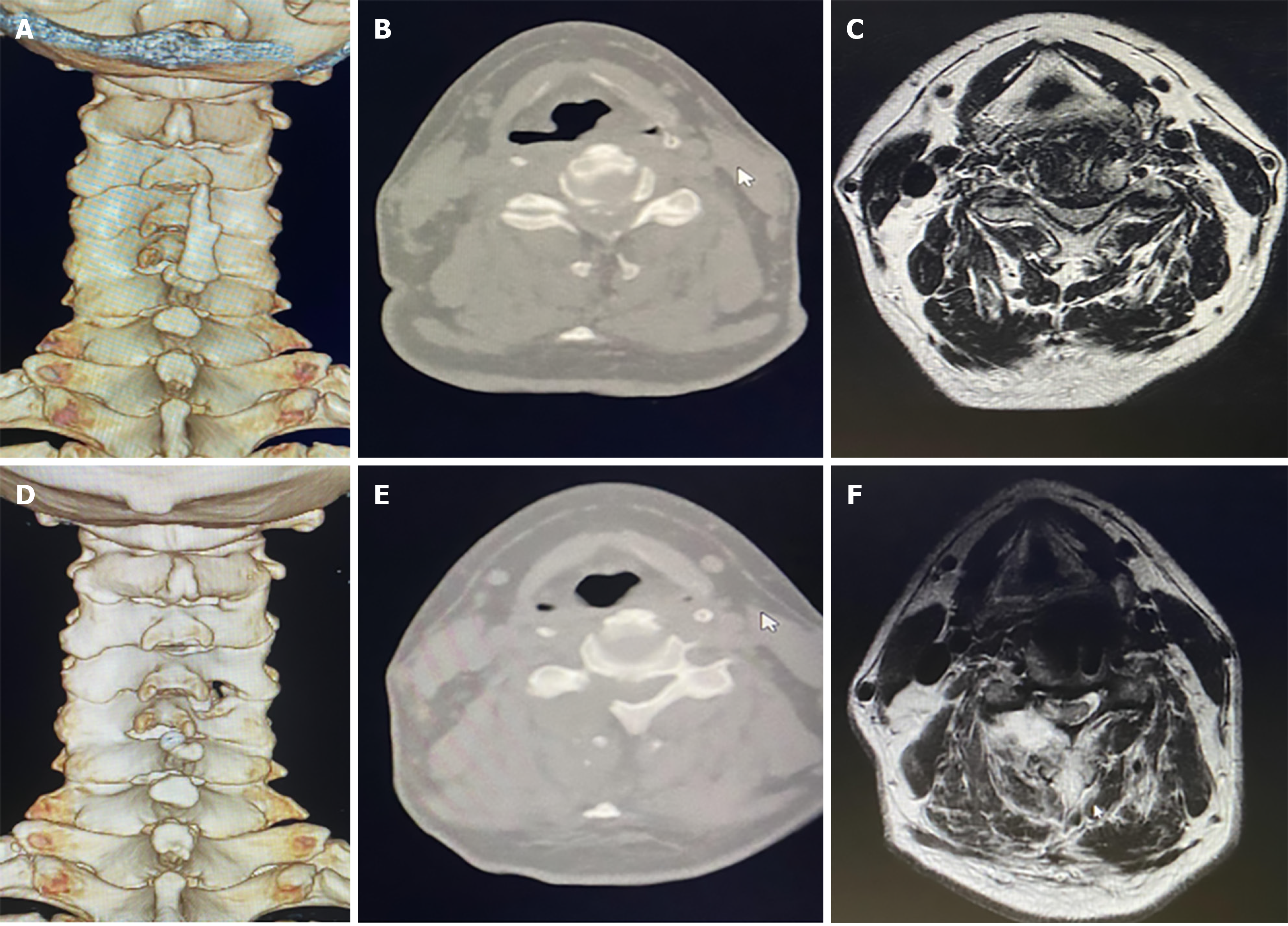
Figure 3 Comparison of pre- and post-operative computed tomography and magnetic resonance imaging findings.
A: Pre-operative 3D reconstruction; B and C: Pre-operative computed tomography (CT) and magnetic resonance imaging (MRI) showed herniated disc in C4/5; D: Post-operative 3D reconstruction showed keyhole laminotomy at right side in C4/5; E and F: Post-operative CT and MRI showed that nerve root was fully decompressed.
- Citation: Yan MJ, Zhang BT, Tang GK, Liu YB, Liao WB, Guo S, Fu Q. Novel endoscopic hybrid technique in the treatment of cervical spondylotic radiculopathy. World J Orthop 2025; 16(8): 109904
- URL: https://www.wjgnet.com/2218-5836/full/v16/i8/109904.htm
- DOI: https://dx.doi.org/10.5312/wjo.v16.i8.109904


