Copyright
©The Author(s) 2025.
World J Orthop. Aug 18, 2025; 16(8): 109904
Published online Aug 18, 2025. doi: 10.5312/wjo.v16.i8.109904
Published online Aug 18, 2025. doi: 10.5312/wjo.v16.i8.109904
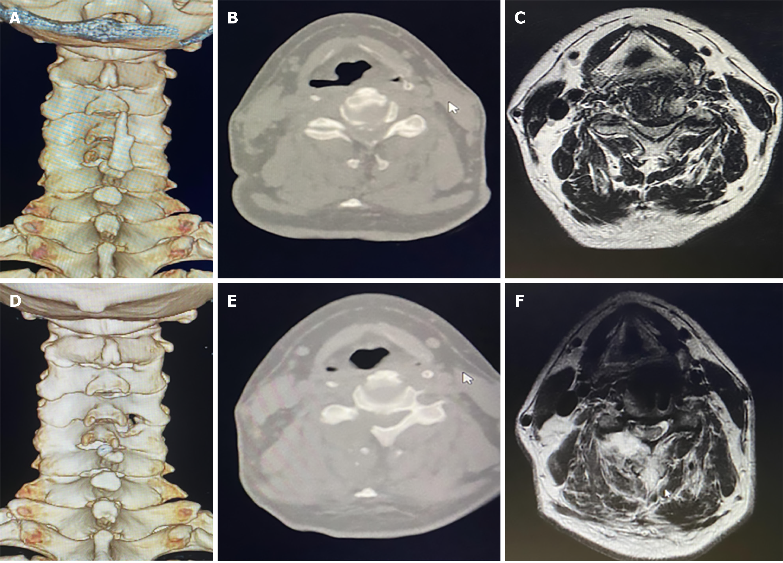
Figure 3 Comparison of pre- and post-operative computed tomography and magnetic resonance imaging findings.
A: Pre-operative 3D reconstruction; B and C: Pre-operative computed tomography (CT) and magnetic resonance imaging (MRI) showed herniated disc in C4/5; D: Post-operative 3D reconstruction showed keyhole laminotomy at right side in C4/5; E and F: Post-operative CT and MRI showed that nerve root was fully decompressed.
- Citation: Yan MJ, Zhang BT, Tang GK, Liu YB, Liao WB, Guo S, Fu Q. Novel endoscopic hybrid technique in the treatment of cervical spondylotic radiculopathy. World J Orthop 2025; 16(8): 109904
- URL: https://www.wjgnet.com/2218-5836/full/v16/i8/109904.htm
- DOI: https://dx.doi.org/10.5312/wjo.v16.i8.109904


