Copyright
©The Author(s) 2025.
World J Radiol. Jun 28, 2025; 17(6): 107522
Published online Jun 28, 2025. doi: 10.4329/wjr.v17.i6.107522
Published online Jun 28, 2025. doi: 10.4329/wjr.v17.i6.107522
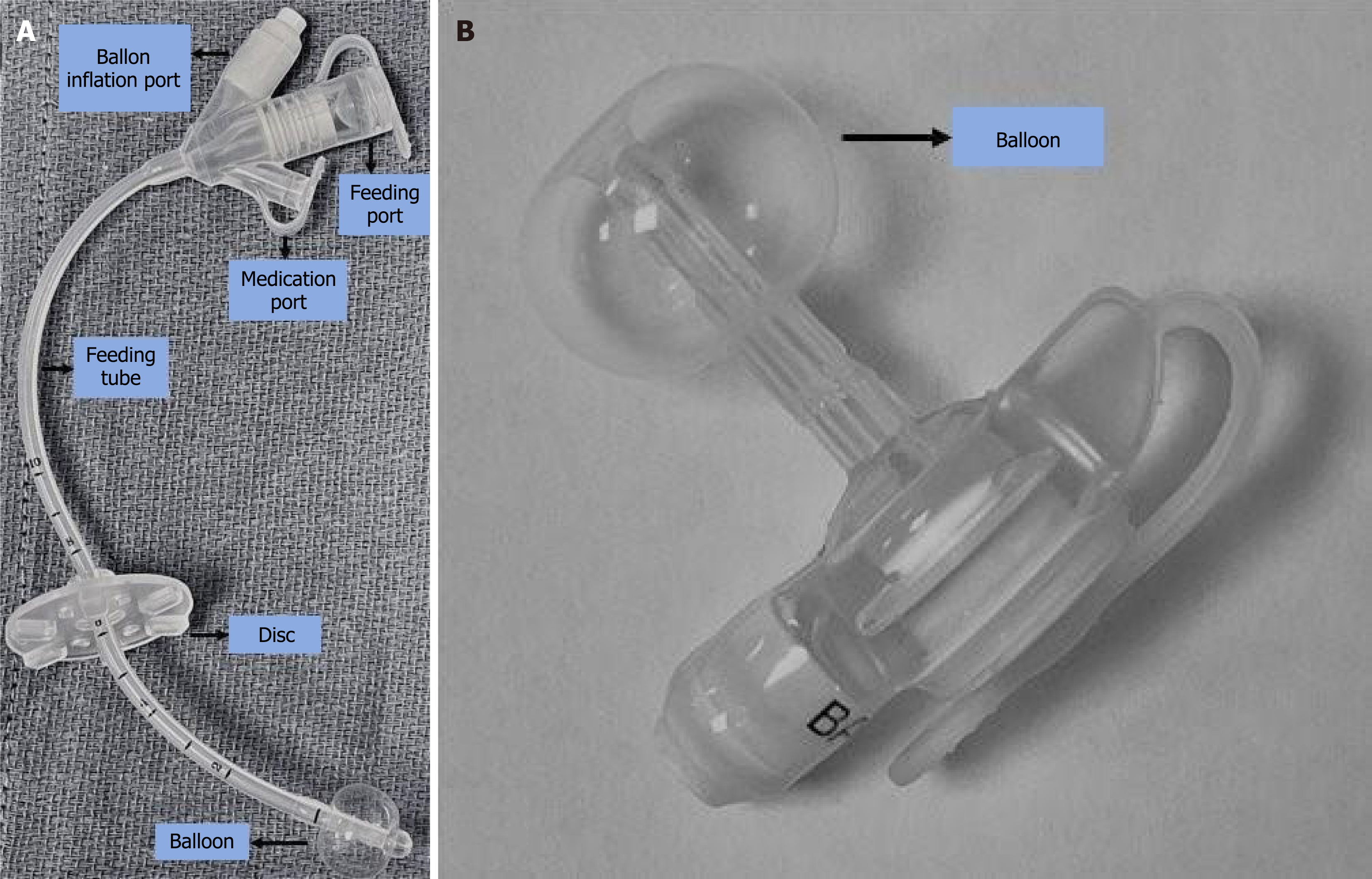
Figure 1 Tube.
A: Standard gastrostomy tube (G-tube); B: Low profile G-tube/ gastrostomy-button.
Figure 2 Normal contrast abdominal radiography study for gastrostomy tube.
A: Frontal; B: Cross table lateral post-injection radiographs show normal outlining of the gastric contour ( white arrows) and rugal folds (arrowheads). There is normal drainage of contrast into the duodenum (curved arrow). No extraluminal spillage noted; C: It is a schematic diagram depicting normal findings which should be looked at including filling defects of gastric rugae in the contrast pool (black arrow), more importantly the filling defect of the gastrostomy tube ballon, gastric contour and tracking of contrast into the duodenum (black arrowhead).
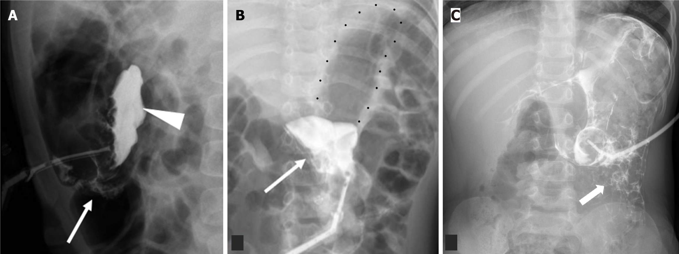
Figure 3 Free peritoneal spillage of contrast.
A: Lateral radiograph of a 1-year-old with recently exchanged tube. Injected contrast pools in the intraperitoneal cavity (arrowhead) which does not follow the stomach contour with outlining of bowel loops (arrow); B: Frontal radiograph irregular or angular margins of the injected contrast (arrow) (multiple black dots outlining the air filled stomach); C: Frontal radiograph of another 4-year-old boy demonstrates similar extrinsic outlining of bowel loops.
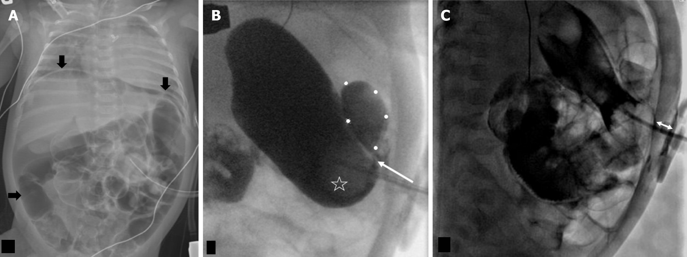
Figure 4 Intraperitoneal leak via the gastrostomy tube tract, 2 month old postoperative day 1 open gastrostomy tube placement.
A: Frontal abdominal radiograph with a large amount of pneumoperitoneum (black arrows); B: Frontal projection in a fluoroscopy gastrostomy tube (G-tube) study with a loculated intraperitoneal leak (outlined by white dotes). There is transit of contrast behind the G-tube ballon (star) into the peritoneal leak (arrow); C: G-tube was exchanged in interventional radiology, following which a lateral fluoroscopic image showed the narrowed distance (double arrow) between the disc and ballon with no trickling of contrast behind it.
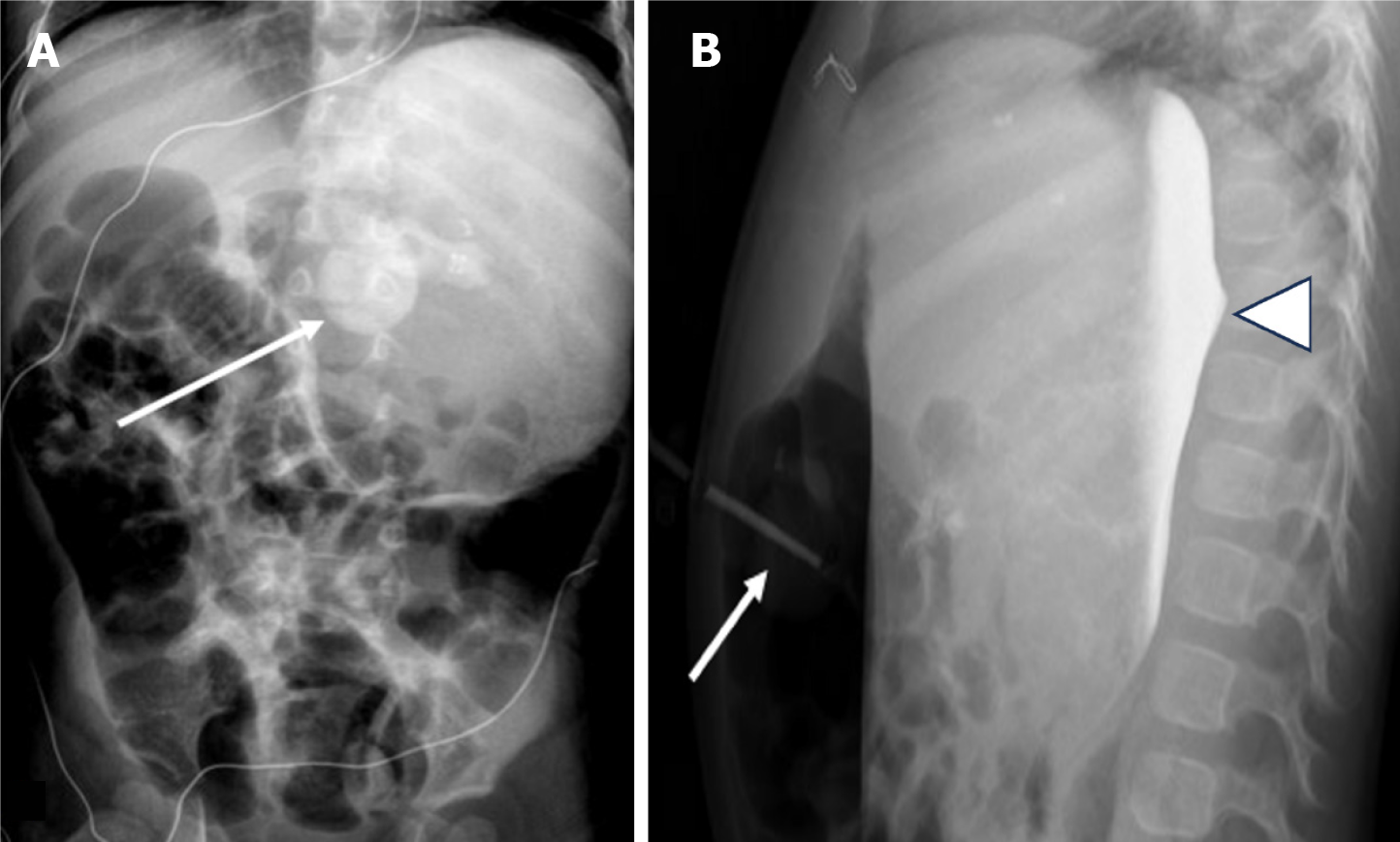
Figure 5 Gastric outlet obstruction secondary to gastrostomy tube ballon.
A: Frontal; B: Lateral. A 2 year old presented with increased abdominal distension, gagging and reflux from gastrostomy tube (G-tube). Portable radiographs, frontal and lateral after contrast administration revealed retention of contrast in an overdistended stomach (arrowhead) with the G-tube balloon in the distal stomach (arrow). Obstruction was later relieved after the balloon (arrow) was deflated and the tube was exchanged.
Figure 6 Malposition of G-port of gastrojejunostomy tube into the duodenum.
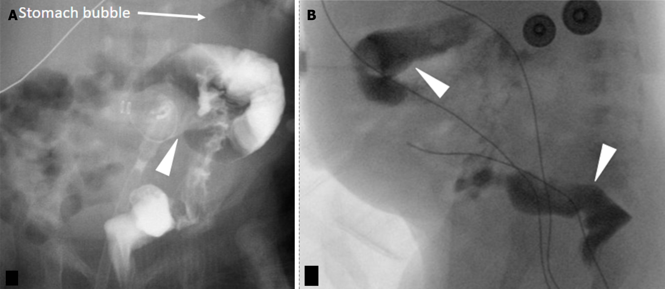
A 20 year old male with increased abdominal distension and bilious output from G-port of gastrojejunostomy tube. A: Frontal radiograph shows that the G-port (black arrow) has migrated into the right upper quadrant from its ideal location in the left upper quadrant suggesting peripyloric position. The G-port should be in the left upper quadrant; B: Oblique post injection radiograph demonstrated intraluminal opacification of the duodenum (arrow) without opacification of the stomach (arrowhead) confirming a migrated G-port balloon into the pylorus or proximal duodenum.
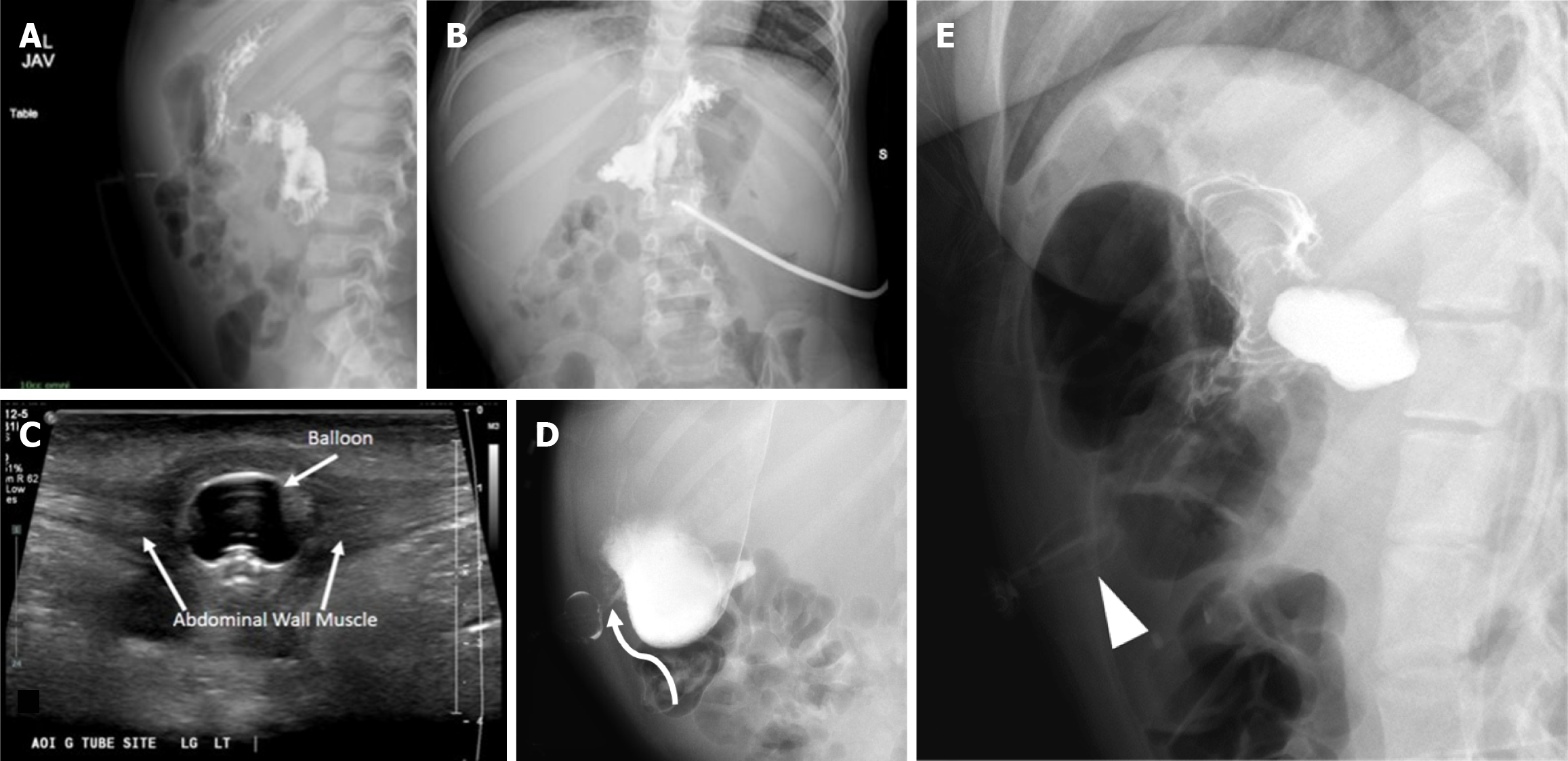
Figure 7 Buried bumper syndrome.
A 5-year-old kid presented with drainage and pain at the gastrostomy tube (G-tube) site. A and B: Contrast abdominal radiography shows contrast filling the stomach and small bowel without extravasation. However, the balloon is not well seen; C: A local site ultrasound revealed G-tube balloon in the anterior abdominal wall along the tract outside of the stomach; D: A follow up fluoroscopy shows delineation of the inflated balloon which lies in the anterior abdominal wall (curved arrow); E: A different case demonstrates a deformed G-tube balloon (arrowhead) in the anterior abdominal wall.
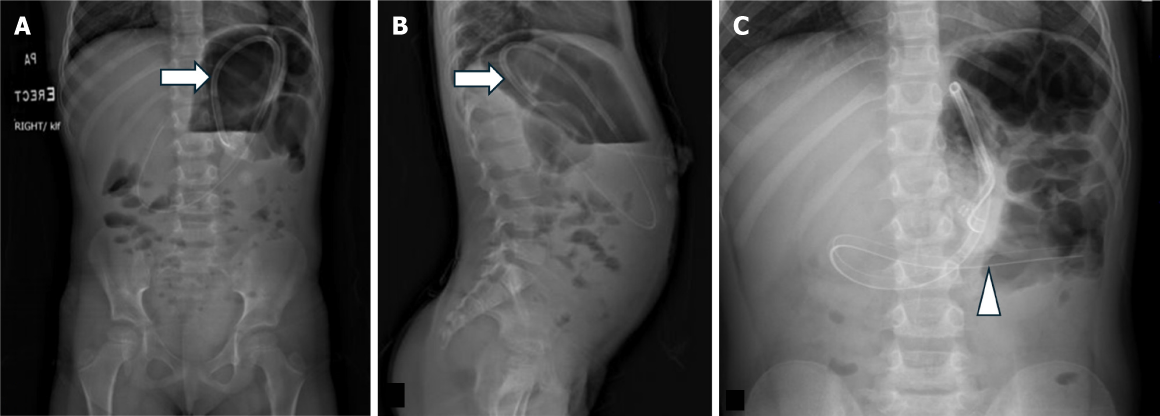
Figure 8 Retraction of gastrojejunostomy tube into the stomach identified incidentally on scoliosis radiographs.
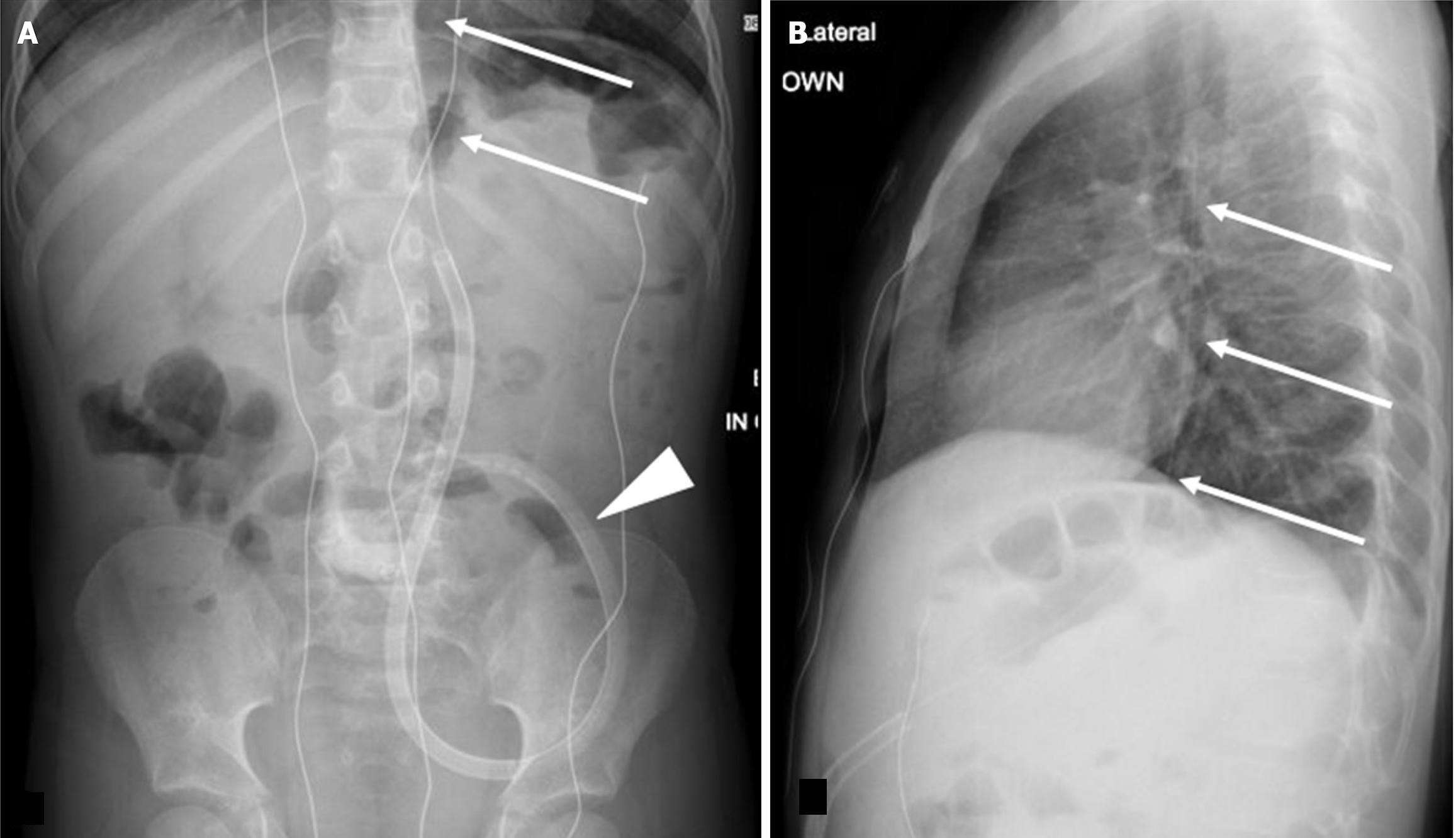
A: Frontal; B: Lateral spine radiographs. They show retraction and coiling of the gastrojejunostomy tube (arrows) into the stomach without a retroperitoneal course; C: Frontal abdominal radiographs of the same patient 10 days prior demonstrates gastrostomy tube coursing normally along the C-loop of the duodenum (arrowhead).
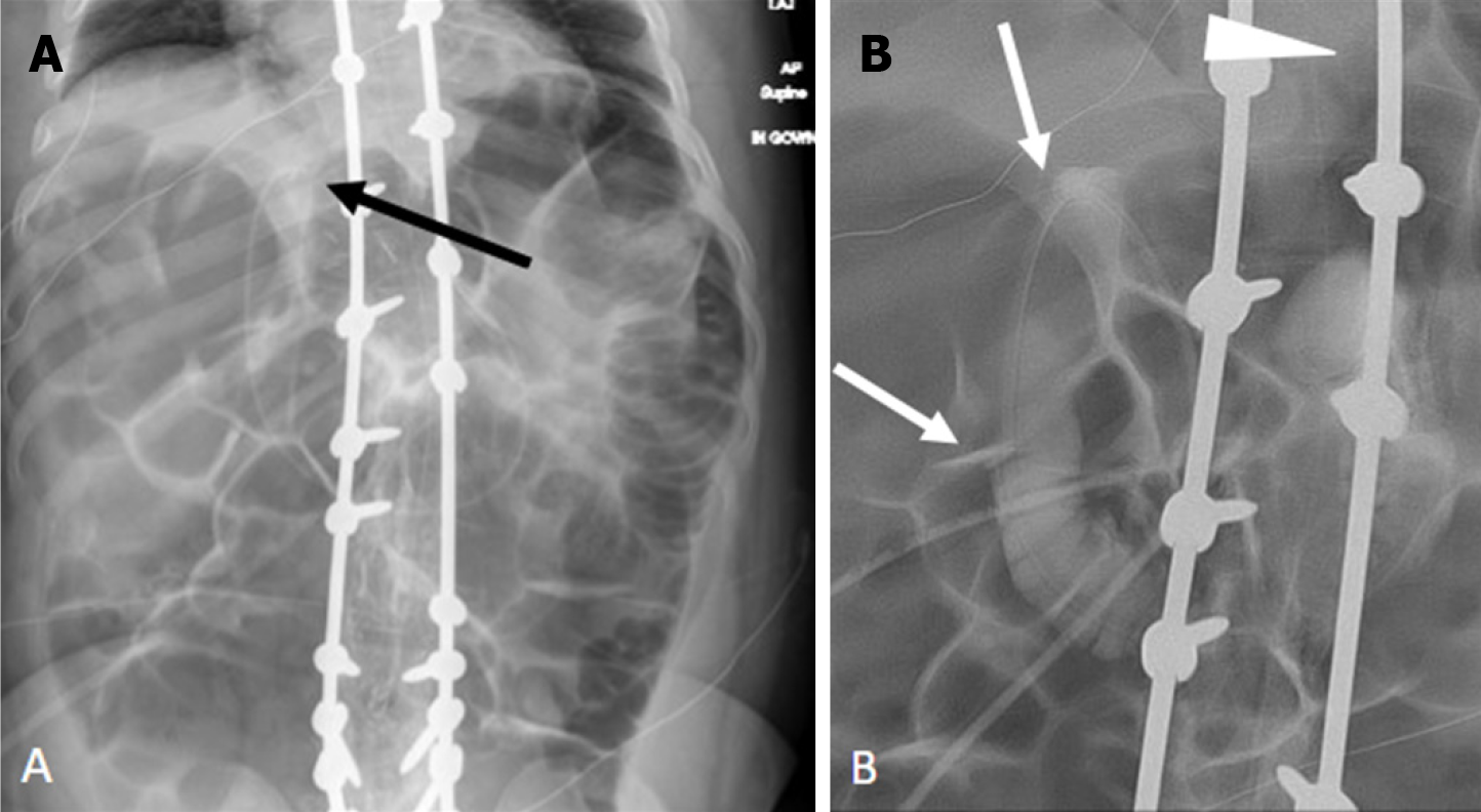
Figure 9 A 16 year old with malfunctioning gastrojejunostomy tube and leakage at stoma.
A: Lateral radiograph depicts a focal discontinuity in the gastrojejunostomy tube, just adjacent to the disc (long arrow); B: Frontal radiographs, pre contrast injection; C: Frontal radiographs, post contrast injection. They depict pooling of contrast at the entry site (short arrow) with no luminal opacification of a jejunal loop. Distal block of the tube could also be an underlying cause leading to tube breakage.
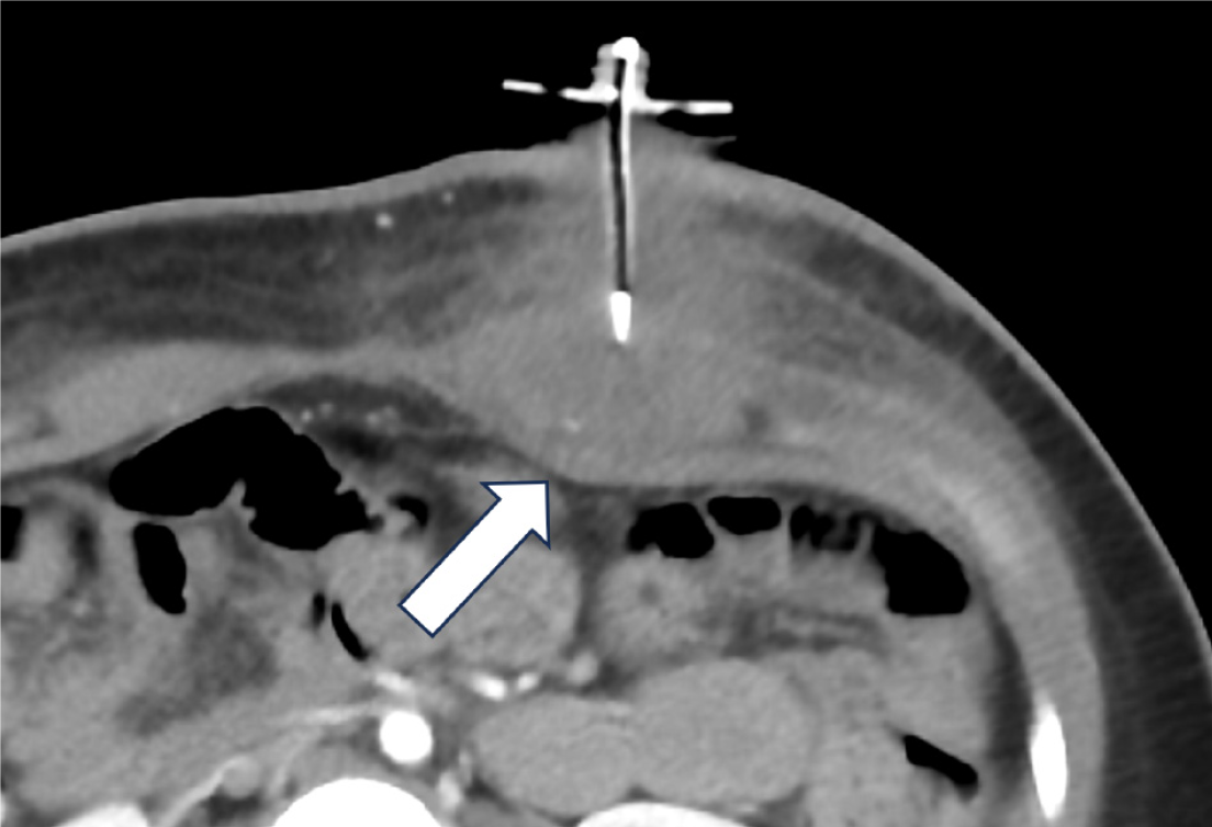
Figure 10 Local site infection.
A 19 year old girl whose gastrostomy button (G-button) was changed at home with presents with a new tube discomfort and swelling. Patient did not follow up regularly since initial G-button placement. There was interval growth of body habitus. A computed tomography was performed on presentation which shows extensive abdominal wall inflammation and inward bowing of abdominal wall (arrow), likely sequela of buried bumper syndrome and leakage of feeds into abdominal wall.
Figure 11 Colonic gastrostomy tube placement.
A 1 month old, postoperative day 5, with a thick brown and bilious output around gastrostomy tube. A and B: Injected contrast demonstrates intraluminal opacification of the transverse colon, descending colon, and rectum (arrowheads) indicating colonic tube placement.
Figure 12 Abnormal gastrostomy tube placement into the bowel loops.
A 16 year old male post operative day 4 percutaneous endoscopic gastrostomy. A and B: Axial sections of the computed tomography abdomen depict the gastrostomy tube transversing the dilated small bowel (arrow) and decompressed transverse colon (arrowhead). Findings were confirmed intraoperatively along with a volvulus.
Figure 13 Esophageal malposition of the gastrojejunostomy tube.
An 8-year-old female with vomiting and balloon visualized in mouth. Due to respiratory concerns, the balloon was deflated and tubing pulled back prior to imaging. A: Abdomen; B: Lateral radiograph of the chest. Anteroposterior radiograph of the abdomen and lateral radiograph of the chest show that the jejunal tubing is in the esophagus (arrows) and gastric tubing (arrowhead) is coiled in a distended stomach.
Figure 14 Local abscess.
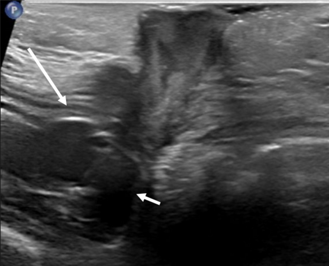
A 9 year old with gastrostomy tube (G-tube) site swelling. Ultrasound shows a complex fluid collection/abscess in the subcutaneous soft tissues. The gastric wall (long arrow) and G-tube balloon (short arrow) are well seen without evidence of intraperitoneal fluid or air.
- Citation: Patel DD, Schenker KE, Averill LW, May LA. Imaging of pediatric gastrostomy tube malposition: Pearls and pitfalls. World J Radiol 2025; 17(6): 107522
- URL: https://www.wjgnet.com/1949-8470/full/v17/i6/107522.htm
- DOI: https://dx.doi.org/10.4329/wjr.v17.i6.107522


