Copyright
©The Author(s) 2025.
World J Radiol. Jun 28, 2025; 17(6): 107522
Published online Jun 28, 2025. doi: 10.4329/wjr.v17.i6.107522
Published online Jun 28, 2025. doi: 10.4329/wjr.v17.i6.107522
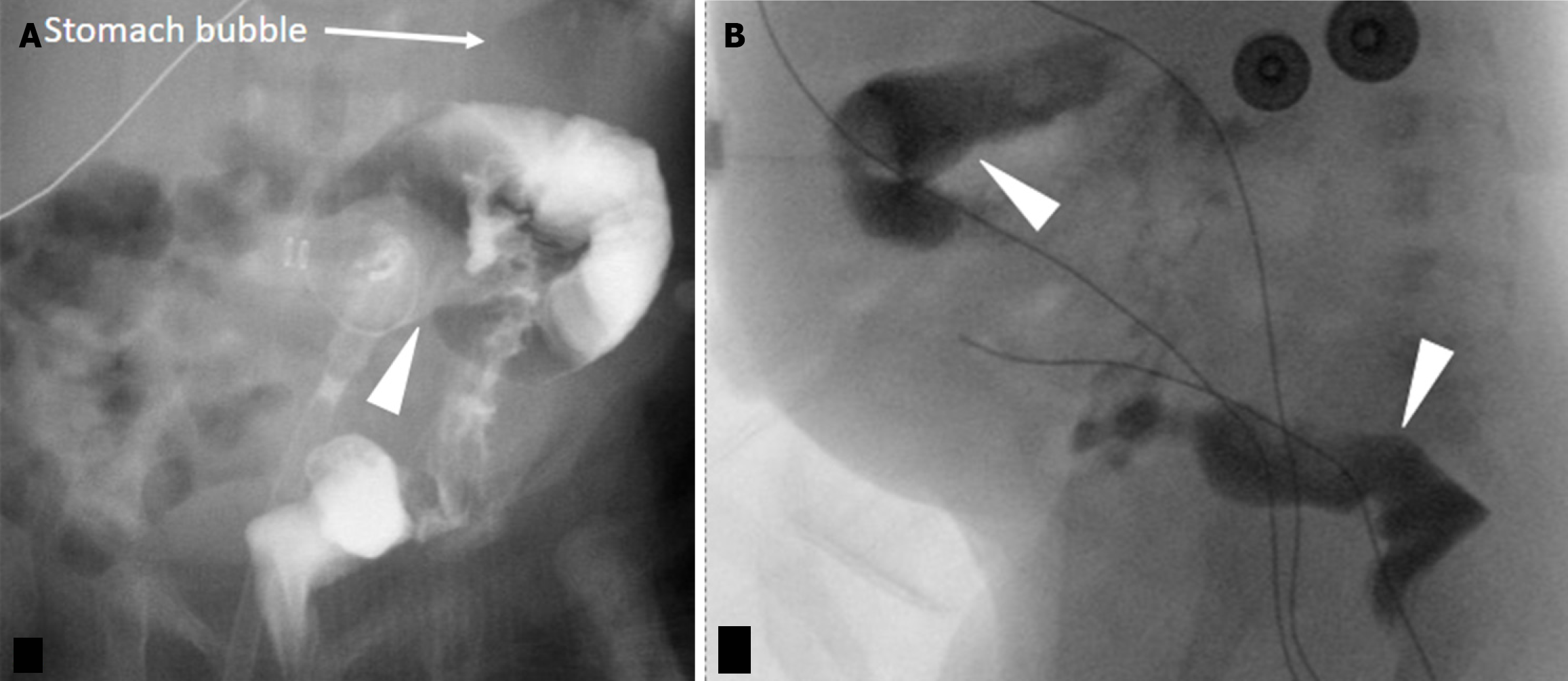
Figure 11 Colonic gastrostomy tube placement.
A 1 month old, postoperative day 5, with a thick brown and bilious output around gastrostomy tube. A and B: Injected contrast demonstrates intraluminal opacification of the transverse colon, descending colon, and rectum (arrowheads) indicating colonic tube placement.
- Citation: Patel DD, Schenker KE, Averill LW, May LA. Imaging of pediatric gastrostomy tube malposition: Pearls and pitfalls. World J Radiol 2025; 17(6): 107522
- URL: https://www.wjgnet.com/1949-8470/full/v17/i6/107522.htm
- DOI: https://dx.doi.org/10.4329/wjr.v17.i6.107522


