Copyright
©The Author(s) 2025.
World J Radiol. Jun 28, 2025; 17(6): 107522
Published online Jun 28, 2025. doi: 10.4329/wjr.v17.i6.107522
Published online Jun 28, 2025. doi: 10.4329/wjr.v17.i6.107522
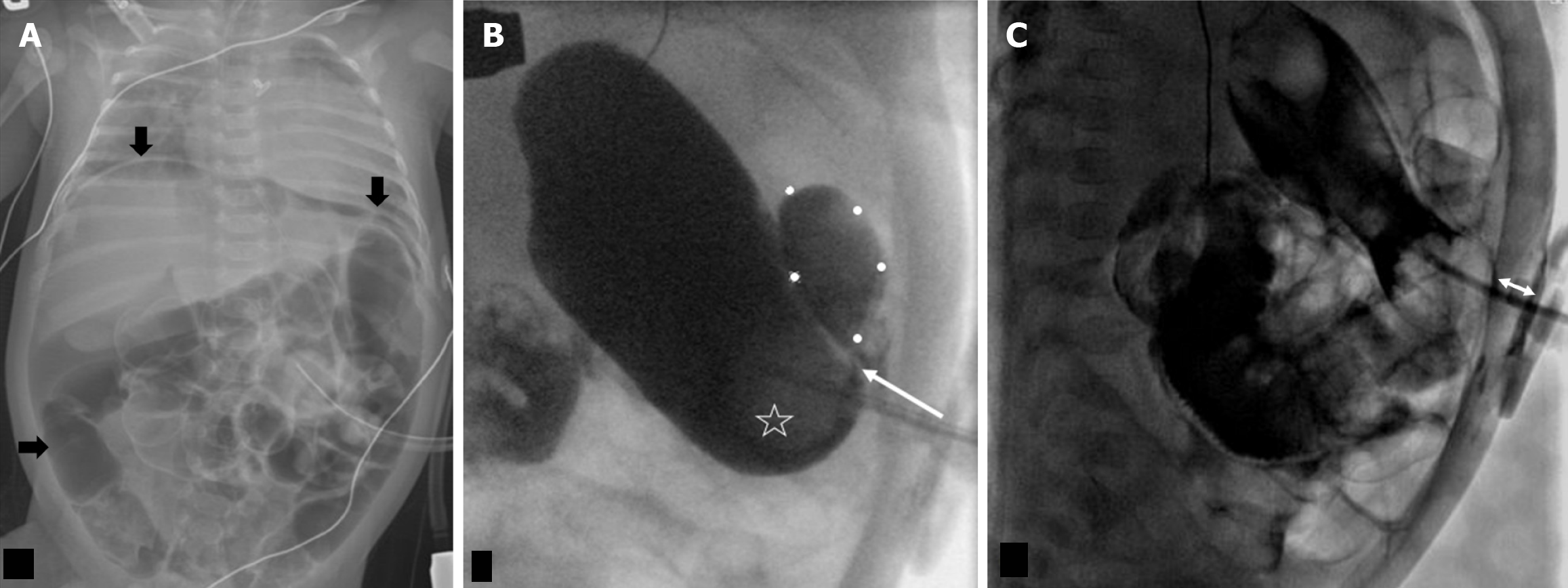
Figure 4 Intraperitoneal leak via the gastrostomy tube tract, 2 month old postoperative day 1 open gastrostomy tube placement.
A: Frontal abdominal radiograph with a large amount of pneumoperitoneum (black arrows); B: Frontal projection in a fluoroscopy gastrostomy tube (G-tube) study with a loculated intraperitoneal leak (outlined by white dotes). There is transit of contrast behind the G-tube ballon (star) into the peritoneal leak (arrow); C: G-tube was exchanged in interventional radiology, following which a lateral fluoroscopic image showed the narrowed distance (double arrow) between the disc and ballon with no trickling of contrast behind it.
- Citation: Patel DD, Schenker KE, Averill LW, May LA. Imaging of pediatric gastrostomy tube malposition: Pearls and pitfalls. World J Radiol 2025; 17(6): 107522
- URL: https://www.wjgnet.com/1949-8470/full/v17/i6/107522.htm
- DOI: https://dx.doi.org/10.4329/wjr.v17.i6.107522


