Copyright
©The Author(s) 2025.
World J Radiol. Jun 28, 2025; 17(6): 107522
Published online Jun 28, 2025. doi: 10.4329/wjr.v17.i6.107522
Published online Jun 28, 2025. doi: 10.4329/wjr.v17.i6.107522
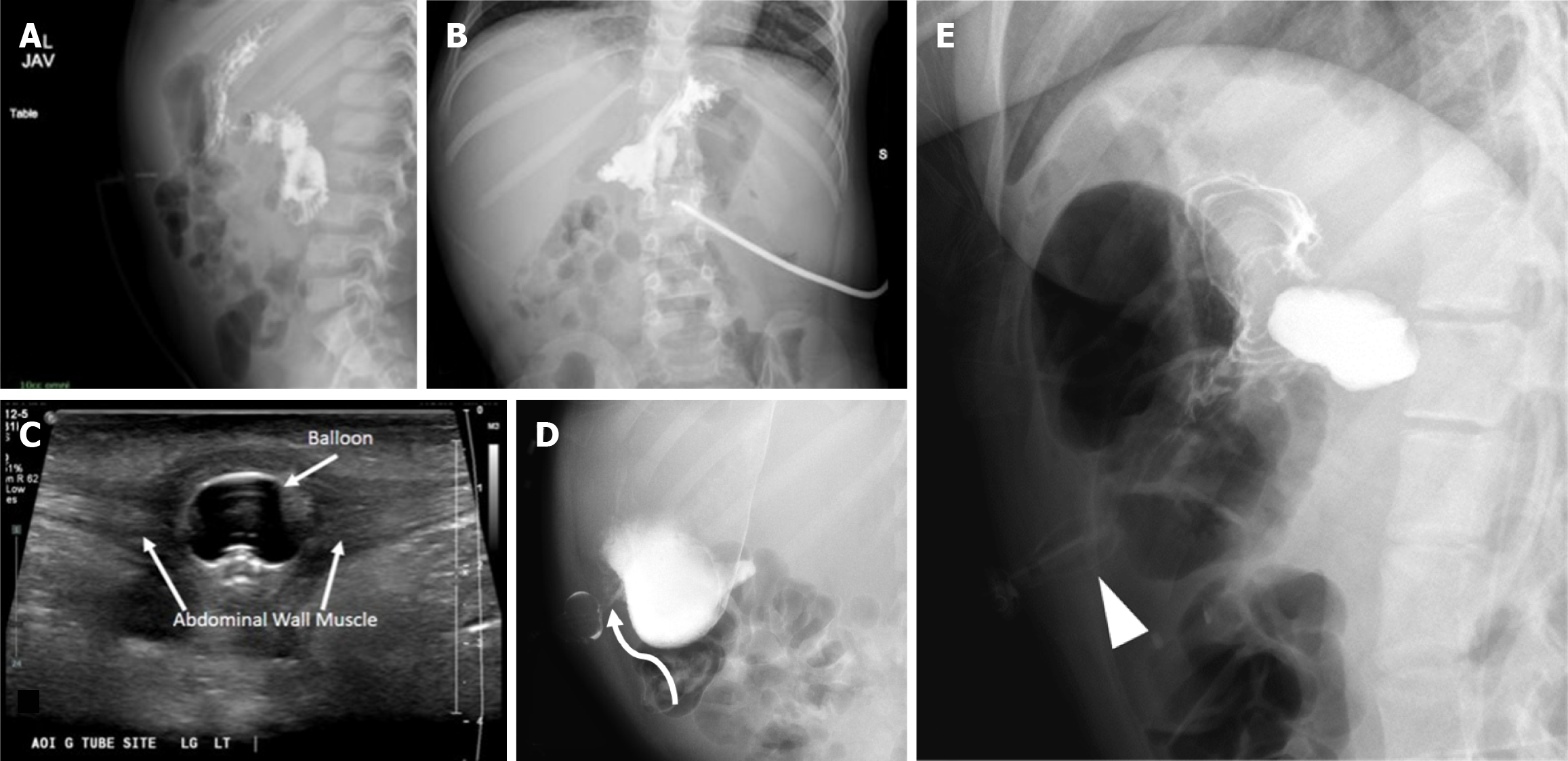
Figure 7 Buried bumper syndrome.
A 5-year-old kid presented with drainage and pain at the gastrostomy tube (G-tube) site. A and B: Contrast abdominal radiography shows contrast filling the stomach and small bowel without extravasation. However, the balloon is not well seen; C: A local site ultrasound revealed G-tube balloon in the anterior abdominal wall along the tract outside of the stomach; D: A follow up fluoroscopy shows delineation of the inflated balloon which lies in the anterior abdominal wall (curved arrow); E: A different case demonstrates a deformed G-tube balloon (arrowhead) in the anterior abdominal wall.
- Citation: Patel DD, Schenker KE, Averill LW, May LA. Imaging of pediatric gastrostomy tube malposition: Pearls and pitfalls. World J Radiol 2025; 17(6): 107522
- URL: https://www.wjgnet.com/1949-8470/full/v17/i6/107522.htm
- DOI: https://dx.doi.org/10.4329/wjr.v17.i6.107522


