Copyright
©The Author(s) 2025.
World J Gastrointest Surg. Jun 27, 2025; 17(6): 107086
Published online Jun 27, 2025. doi: 10.4240/wjgs.v17.i6.107086
Published online Jun 27, 2025. doi: 10.4240/wjgs.v17.i6.107086
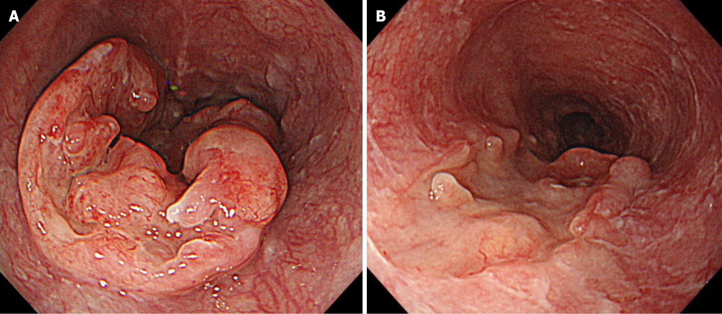
Figure 1 Esophagogastroduodenoscopy findings of the esophageal primary lesion.
A: Imaging at initial presentation reveals a semicircular type 3 tumor in the middle thoracic esophagus, located 29-36 cm away from the incisors. Histological analysis confirmed squamous cell carcinoma; B: After three courses of docetaxel + cisplatin + 5-fluorouracil chemotherapy, imaging revealed significant shrinkage of the primary lesion.
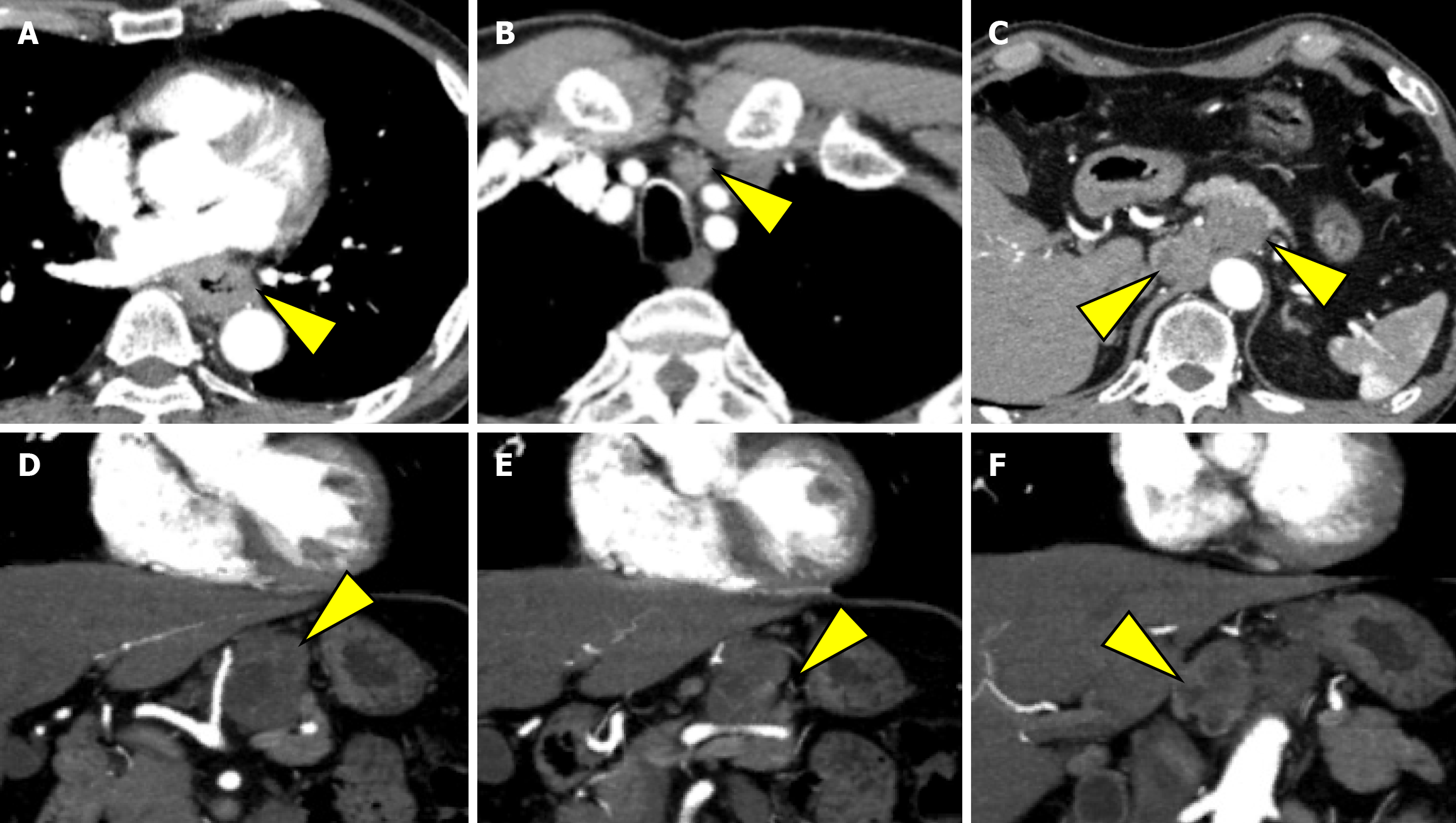
Figure 2 Computed tomography findings at the initial presentation.
A: Image showing irregular wall thickening from the middle to the lower thoracic esophagus as the primary lesion; B: Imaging showing enlarged No. 101L lymph node measuring 15 mm; C: Imaging showing bulky suprapancreatic lymph node metastasis, including No. 8, 9, and 11p; D and E: Imaging showing enlargement of the No. 11p lymph node to 35 mm, directly invading the pancreatic body; F: Imaging showing enlarged No. 8 and No. 9 lymph nodes, forming a mass.
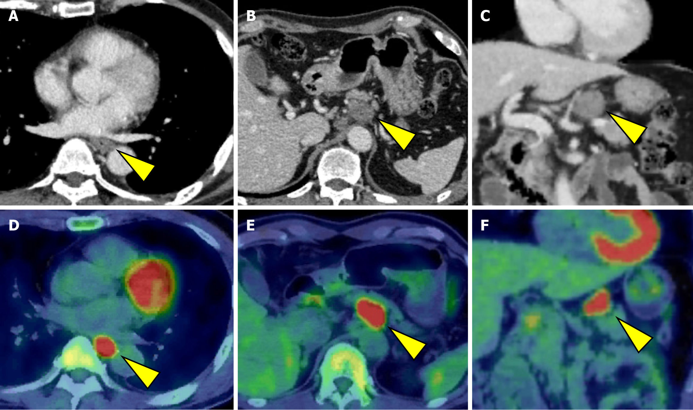
Figure 3 Computed tomography and positron emission tomography-computed tomography findings, three courses after docetaxel + cisplatin + 5-fluorouracil chemotherapy.
A: Imaging showing improved irregular wall thickening at the primary lesion; B and C: Imaging showing a remarkable reduction in the sizes of metastases of lymph nodes 8 and 9. However, the No. 11p lymph node had shrunk slightly, and the border with the pancreas appeared to have improved indistinctiveness; D: Fluorodeoxyglucose accumulation in the primary lesion (SUVmax 28.95); E and F: Fluorodeoxyglucose accumulation in No. 11p lymph node metastasis (SUVmax 20.77).
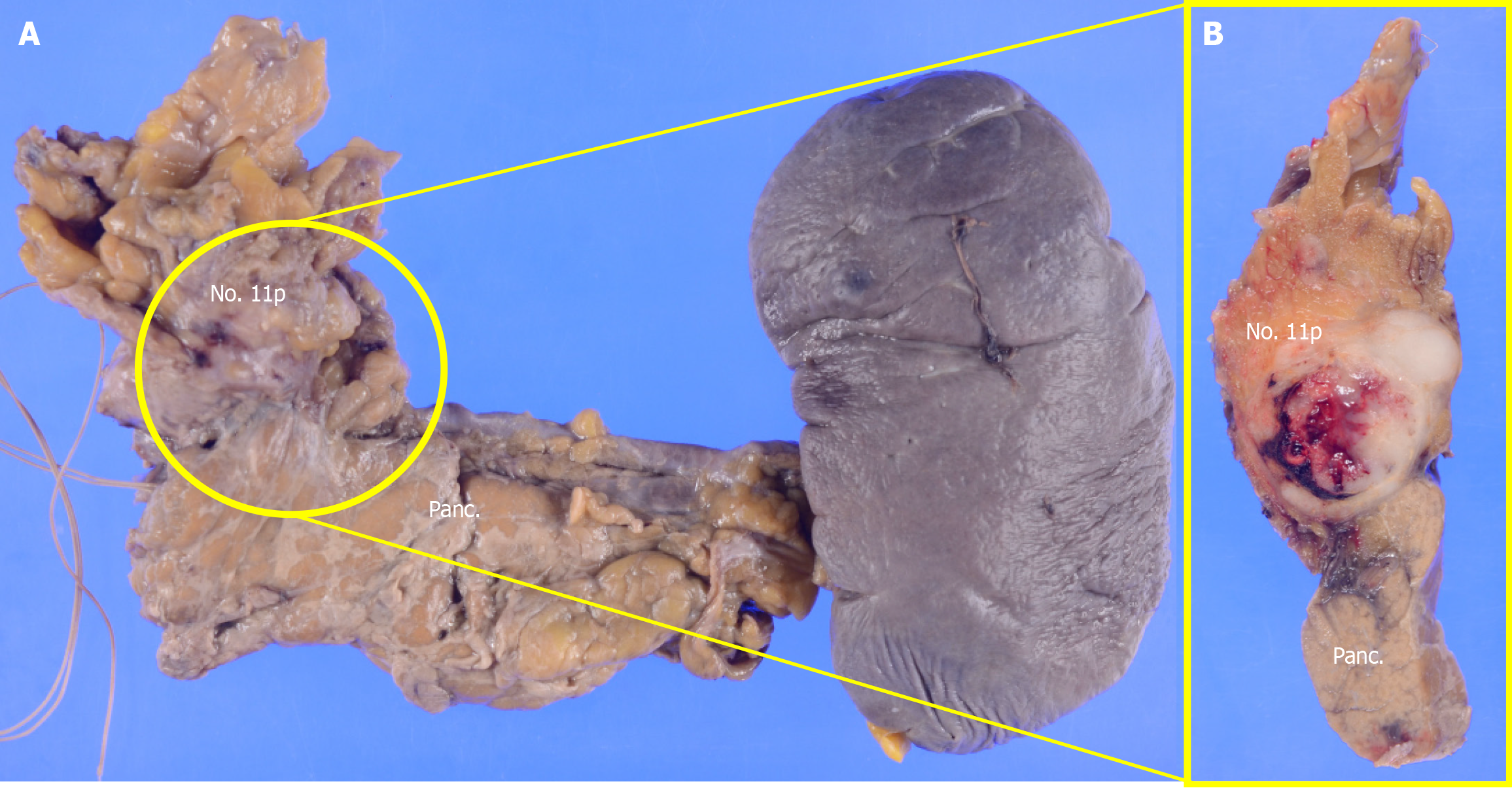
Figure 4 Gross findings of the resected specimen.
A and B: The resected pancreatic body and spleen showed that the No. 11p lymph node was macroscopically attached to the pancreas; however, pathological examination confirmed the absence of direct infiltration. Panc: Pancreas.
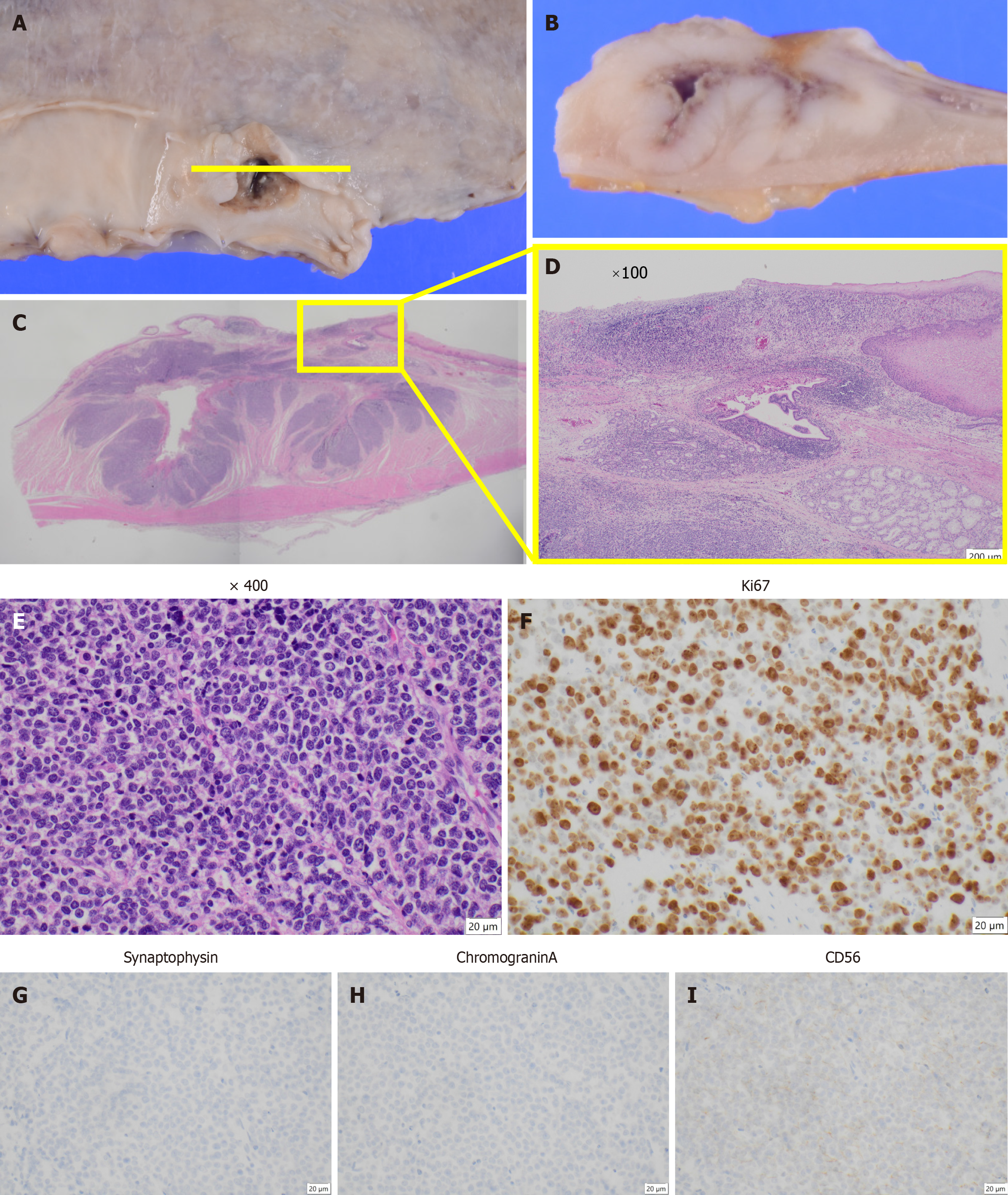
Figure 5 Histological and immunohistochemical findings.
A and B: Type 3 tumor located in the middle thoracic esophagus with the depth of invasion classified as T2 (muscularis propria); C: Cross-sectional hematoxylin and eosin staining showing the tumor structure; D: Magnified view of the area indicated by the square in panel C; E: Hematoxylin and eosin staining reveals a large-cell type of neuroendocrine carcinoma, with no coexistence of squamous cell carcinoma at × 400 magnification; F: Ki-67 staining was positive in more than 90% of tumor cells, indicating a high proliferative index; G-I: Immunohistochemical analysis showed that the tumor stained positively for CD56, but not synaptophysin or chromogranin A.
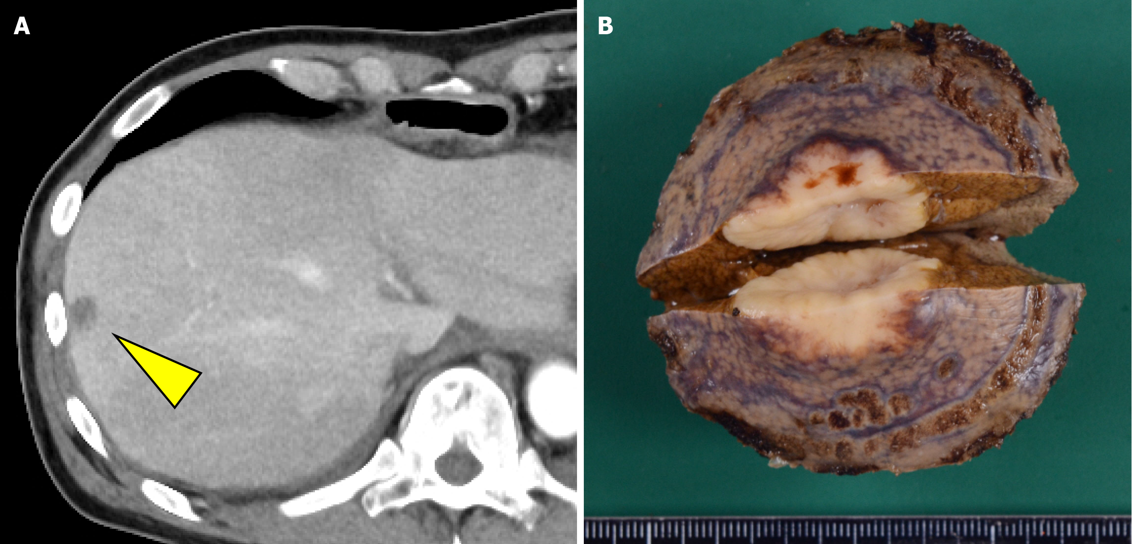
Figure 6 Imaging of the metastasis to the liver segment 8.
A: Computed tomography performed within one month postoperatively revealed a hypovascular nodule in liver segment 8; B: Gross finding of resected liver segment 8.
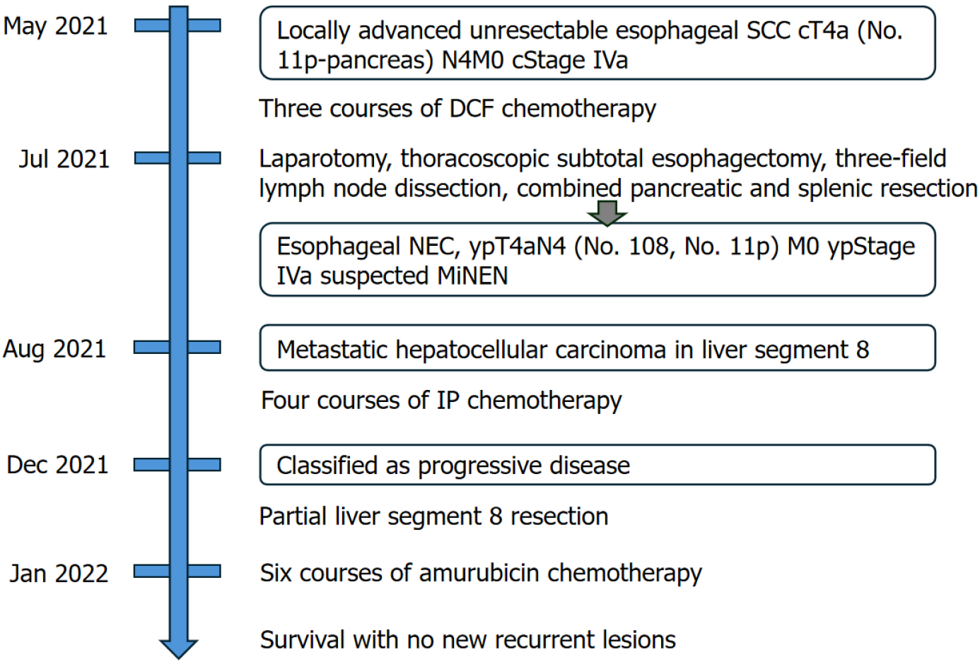
Figure 7 Summary of the treatment course.
SCC: Squamous cell carcinoma; DCF: Docetaxel, cisplatin, and 5-fluorouracil; NEC: Neuroendocrine carcinoma; MiNEN: Mixed neuroendocrine non-neuroendocrine neoplasm; IP: Irinotecan + cisplatin.
- Citation: Okamoto K, Fujisawa K, Kono K, Ogawa Y, Shimoyama H, Haruta S, Takazawa Y, Ueno M, Udagawa H. Long-term survival with multimodal treatment including conversion surgery for locally advanced esophageal neuroendocrine carcinoma: A case report. World J Gastrointest Surg 2025; 17(6): 107086
- URL: https://www.wjgnet.com/1948-9366/full/v17/i6/107086.htm
- DOI: https://dx.doi.org/10.4240/wjgs.v17.i6.107086


