Copyright
©The Author(s) 2025.
World J Gastrointest Surg. Jun 27, 2025; 17(6): 107086
Published online Jun 27, 2025. doi: 10.4240/wjgs.v17.i6.107086
Published online Jun 27, 2025. doi: 10.4240/wjgs.v17.i6.107086
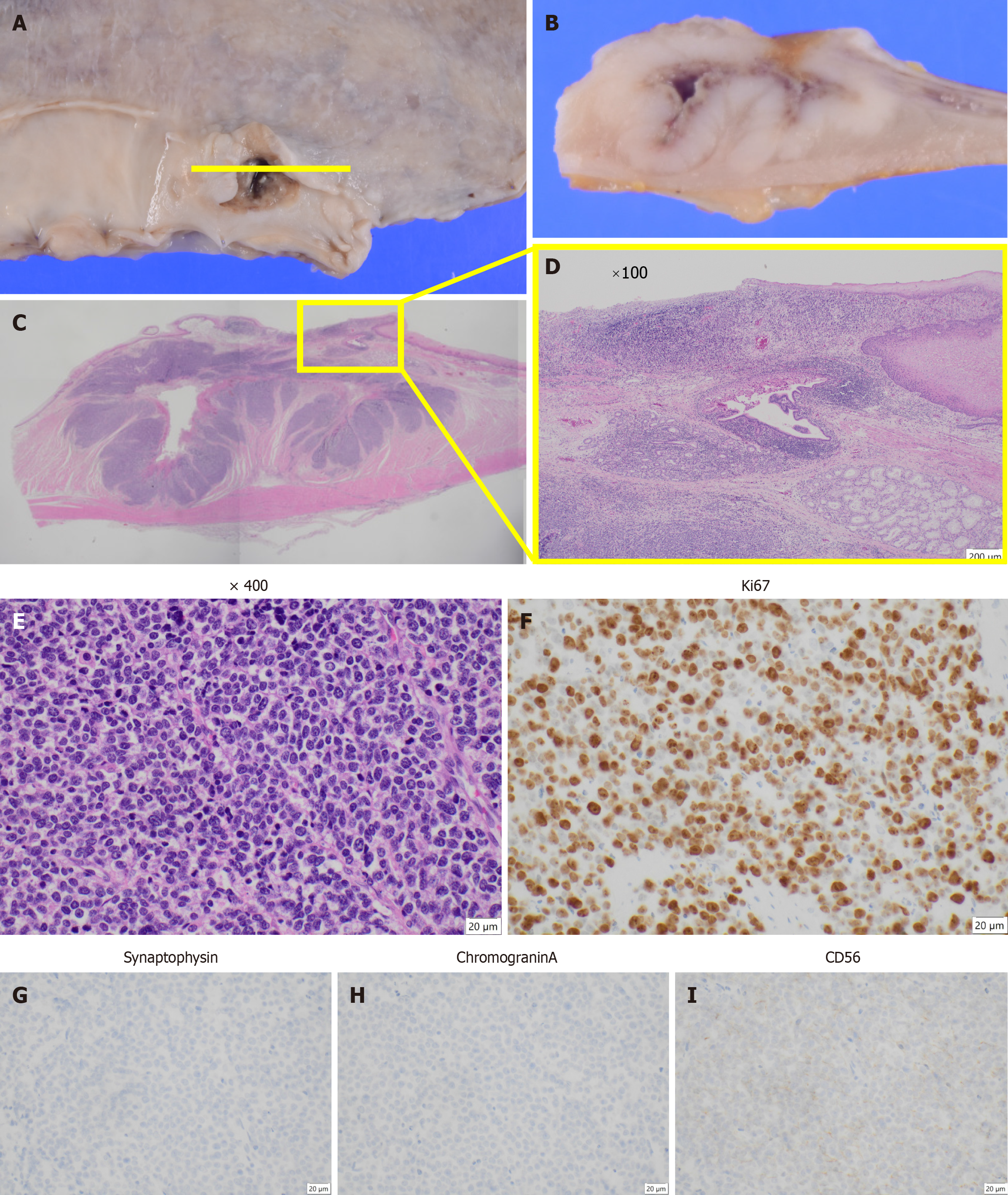
Figure 5 Histological and immunohistochemical findings.
A and B: Type 3 tumor located in the middle thoracic esophagus with the depth of invasion classified as T2 (muscularis propria); C: Cross-sectional hematoxylin and eosin staining showing the tumor structure; D: Magnified view of the area indicated by the square in panel C; E: Hematoxylin and eosin staining reveals a large-cell type of neuroendocrine carcinoma, with no coexistence of squamous cell carcinoma at × 400 magnification; F: Ki-67 staining was positive in more than 90% of tumor cells, indicating a high proliferative index; G-I: Immunohistochemical analysis showed that the tumor stained positively for CD56, but not synaptophysin or chromogranin A.
- Citation: Okamoto K, Fujisawa K, Kono K, Ogawa Y, Shimoyama H, Haruta S, Takazawa Y, Ueno M, Udagawa H. Long-term survival with multimodal treatment including conversion surgery for locally advanced esophageal neuroendocrine carcinoma: A case report. World J Gastrointest Surg 2025; 17(6): 107086
- URL: https://www.wjgnet.com/1948-9366/full/v17/i6/107086.htm
- DOI: https://dx.doi.org/10.4240/wjgs.v17.i6.107086


