Copyright
©The Author(s) 2025.
World J Gastrointest Surg. Jun 27, 2025; 17(6): 107086
Published online Jun 27, 2025. doi: 10.4240/wjgs.v17.i6.107086
Published online Jun 27, 2025. doi: 10.4240/wjgs.v17.i6.107086
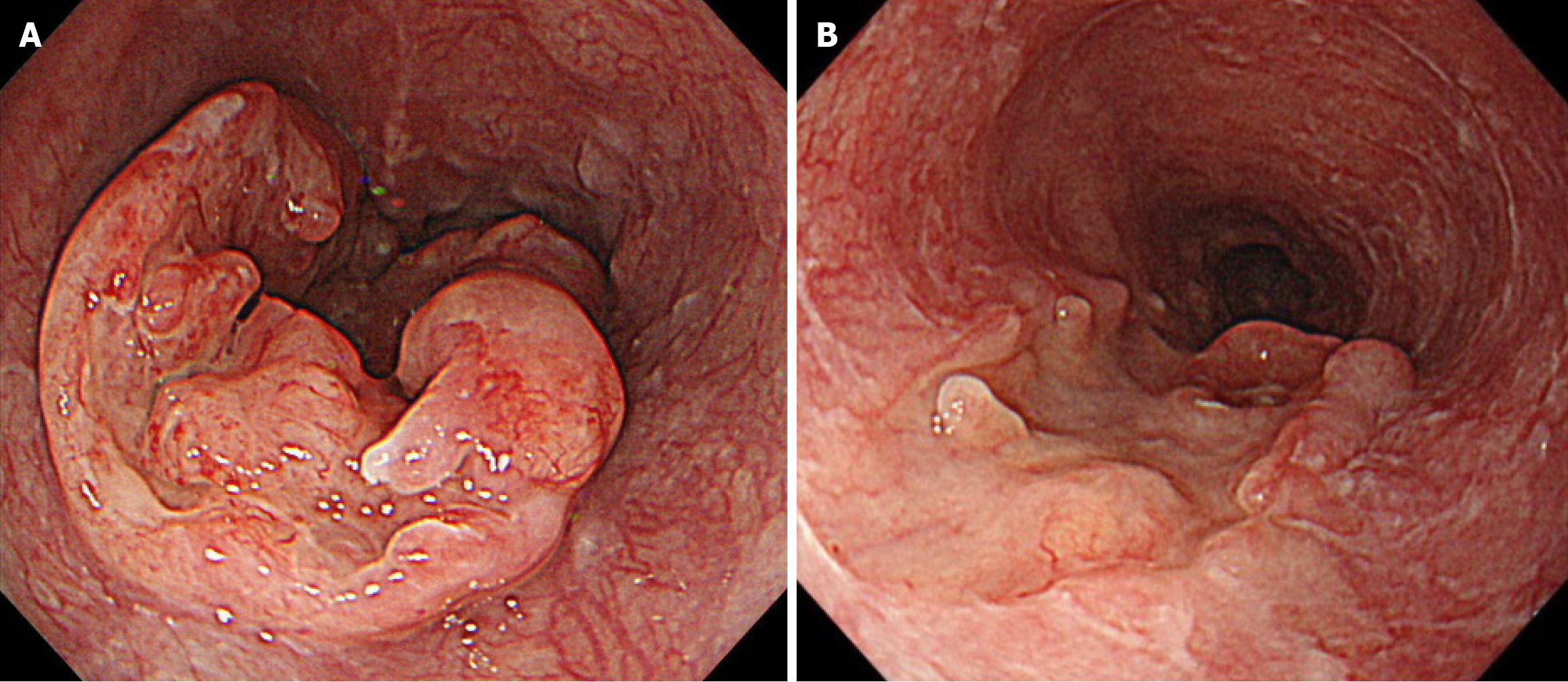
Figure 1 Esophagogastroduodenoscopy findings of the esophageal primary lesion.
A: Imaging at initial presentation reveals a semicircular type 3 tumor in the middle thoracic esophagus, located 29-36 cm away from the incisors. Histological analysis confirmed squamous cell carcinoma; B: After three courses of docetaxel + cisplatin + 5-fluorouracil chemotherapy, imaging revealed significant shrinkage of the primary lesion.
- Citation: Okamoto K, Fujisawa K, Kono K, Ogawa Y, Shimoyama H, Haruta S, Takazawa Y, Ueno M, Udagawa H. Long-term survival with multimodal treatment including conversion surgery for locally advanced esophageal neuroendocrine carcinoma: A case report. World J Gastrointest Surg 2025; 17(6): 107086
- URL: https://www.wjgnet.com/1948-9366/full/v17/i6/107086.htm
- DOI: https://dx.doi.org/10.4240/wjgs.v17.i6.107086


